Download
1 / 31
310 likes | 409 Views
Chapter 17. The Special Senses. Special Senses. Recall that a sensation is the conscious or subconscious awareness of an internal or external stimulus

E N D
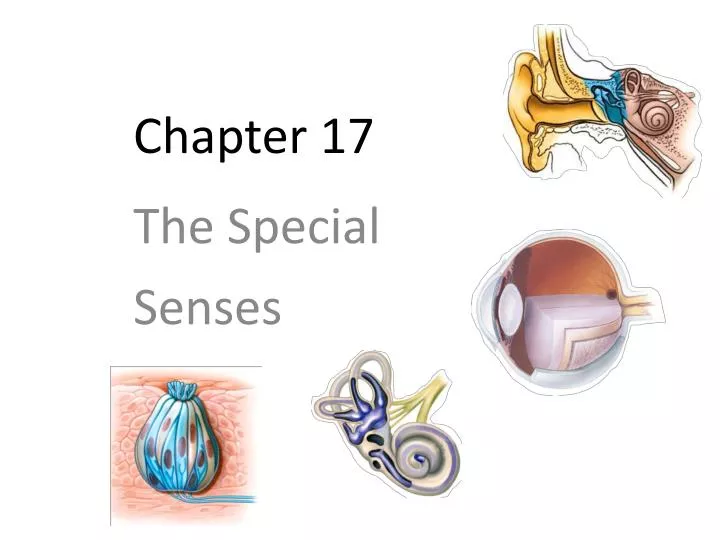
Chapter 17 The Special Senses
Special Senses • Recall that a sensation is the conscious or subconscious awareness of an internal or external stimulus • For this chapter, “external stimulus” means light rays striking the retina of the eye, sound waves impinging on the tympanic membrane of the ear, moleculesin the air and food transmitting smells and tastes to the chemical sensors in the nose an on the tongue, and the force of gravity acting on equilibrium receptors in the inner ear which sense changes in inertia
Special Senses • Receptors for the special senses of smell, taste, vision, hearing, and equilibrium are anatomically distinct from one another and are concentrated in specific locations in the head • In addition to the stimuli and the receptors, there are specific afferent pathways and translation sites in the brain for information assembled from these special senses
Special Senses • Comparing the general senses and the special senses General Senses • Include somatic sensations (tactile, thermal, pain, and proprioceptive) and visceral sensations • Are scattered throughout the body • Are relatively simple structures Special Senses • Include smell, taste, vision, hearing and equilibrium • Are concentrated in specific locations in the head • Are anatomically distinct structures • Form complex neural pathways
Olfaction and Taste • Olfaction is the process of perceiving smells. Smell and taste are brought about through the interpretation of chemicals present in the environment • Olfactory and gustatory (taste) impulses travel not only to the cerebral cortex, but also to the limbic system • this is why we can have emotional responses and trigger strong memories to certain smells and tastes • gustation and olfaction work together but olfaction is much stronger/more sensitive (when someone has a cold it is difficult to taste food)
Olfaction • The olfactory epithelium is located in the superior part of the nasal cavity covering the surface of the cribriform plate and extending along the superior nasal concha
Olfaction • The olfactory epithelium consists of 3 kinds of cells: • The olfactory receptor is a bipolar neuron with cilia (called olfactory hairs). There are 10-100 million of these receptors in the nose that respond to odorant molecules • Supporting cells provide support and nourishment • Basal cells are stem cells that replace olfactory receptors
Olfaction • The olfactory apparatus can detect about 10,000 different odors, often in concentrations as low as 1/25 billionth of a milligram per milliliter of air • When an odorant binds to the receptor of an olfactory hair it initiates a cascade of intracellular events through a G-protein and a 2nd messenger ( production of cAMP opening of Na+ channels inflow of Na+ generator potentials)
Olfaction • Once generated, nerve impulses travel through the two olfactory nerves olfactory bulbs olfactory tract primary olfactory area in the temporal lobe of the cortex • Olfaction is the only sensory system that has direct cortical projections without first going through relay stations in the thalamus
Olfaction • Olfactory sensory pathways (centrally) are rapidly adapting, decreasing activity by 50% in the first second, and completely accommodating in 1–2 minutes • Olfactory supporting cells and glands are innervated by the facial (VII) nerve, a component of which provides parasympathetic motor innervationto lacrimal glands and the mucous membranes in the nasal cavity. This is why certain odors will make our nose run and cause us to produce tears
Gustation • Gustation, or taste, is much simpler than olfaction in that only five primary tastes can be distinguished: sour, sweet, bitter, salty, and umami (“meaty” or “savory”) • Umami is believed to arise from taste receptors that are stimulated by monosodium glutamate (MSG), a substance naturally present in many foods and added to others as a flavor enhancer • All other flavors, such as chocolate, pepper, and coffee, are combinations of the five primary tastes, plus accompanying olfactory and tactile (touch) sensations
Gustation • We have nearly 10,000 taste buds located on the tongue, soft palate, pharynx, and larynx (the number with age) • Each taste bud is composed of about 50 gustatory receptor cells, surrounded by a number of supporting cells • Basal cells located near the CT base multiply and differentiate, first to become the supporting cells around the bud, then the gustatory receptor cells inside the taste bud
Gustation • A single, long microvillus, called a gustatory hair, projects from each receptor cell to the surface through the taste pore • Each gustatory receptor cell has a lifespan of about 10 days
Gustation • Taste buds are found in 3 different types of papillae (elevations on the tongue which provide a rough texture • About 12 very large vallate papillae form a row at the back of the tongue (each houses 100–300 taste buds) • Fungiform papillae are mushroom-shaped and are scattered over the entire surface of the tongue (containing about 5 taste buds each) • Foliate papillae are located in small trenches on the lateral margins of the tongue, but most of their taste buds degenerate in early childhood
Gustation • In addition, the entire surface of the tongue has filiform papillaethat contain tactile receptors but no taste buds • They increase friction between the tongue and food, making it easier to move food in the oral cavity
Gustation • Three cranial nerves contain axons of the first-order gustatory neurons that innervate the taste buds • The facial (VII) nerveserves taste buds in the anterior 2/3 of the tongue • The glossopharyngeal (IX) nerve serves taste buds in the posterior 1/3 of the tongue • The vagus (X) nerve serves taste buds in the throat and epiglottis
Gustation • Nerve impulses propagate along these cranial nerves to the gustatory nucleus in the medulla oblongata. From there, axons carrying taste signals project to the hypothalamus, limbic system, and thalamus • Taste is perceived consciously as signals from the thalamus arrive at the primary gustatory area at the base of the somatosensory cortex in the parietal lobe
Gustation • The threshold for taste varies for each of the primary tastes • We are most sensitive to bitter substances, such as quinine. Because poisonous substances are often bitter, this high sensitivity may have a protective function • The threshold for sour substances is somewhat higher, followed by salty and sweet substances • Complete adaptation to a specific taste can occur in 1–5 minutes of continuous stimulation
Vision • Our visual perception is dependent on the eye, its accessory structures, the optic tracts, and the 1o visual cortex and it’s association areas • Vision is possible because of photoreceptors that are able to “catch” photons of EM radiation in the 400-700 nm wavelengths – what we perceive as visual light
Vision • The eyeball is about 2.5 cm in diameter, with only about 16% of it viewable by just looking at a person • The accessory structures of the eye are the extraocular muscles, palpebra, conjunctiva, and the lacrimal glands and ducts. The pupil is an opening for light to pass into the back of the eye
Accessory Eye Structures • The upper and lower palpebrae are the eyelids, with the fissure being the space between them • CN III supplies 4 of the 6 extraocular muscles, plus the levator palpebrae superioris muscles that raise the upper eyelid • The conjunctiva is a clear mucous membrane that covers the white (avascular) part of the eye
Accessory Eye Structures • The lacrimal glands are each about the size an almond, situated superolateral to the eyeball. Leading from the lacrimal glands are 6 to 12 excretory lacrimal ducts • Tears (lacrimal fluid) run from the lacrimal glands, into the excretory lacrimal ducts, onto the surface of the conjunctiva, over the surface of the eyeball • some lacrimal fluid also evaporates
Accessory Eye Structures • Tears drain into the lacrimal puncta, which are two openings on the nasal side of the extreme edge of the eyeball. Superior and inferior lacrimal canals empty the tears into the nasolacrimal sac and nasolacrimal duct • The right and left sided nasolacrimal ducts empty into each side of the nose
Accessory Eye Structures • Watery eyes occur when lacrimal fluid builds up, as when something obstructs the nasolacrimal ducts for instance • Blocked nasolacrimal ducts can be caused by an inflammation of the nasal mucosa, such as a cold • Over production of lacrimal fluid occurs in response to parasympathetic stimulation, caused by an emotional response (crying), and tears spill over the edges of the eyelids and drain into the nasal cavity (causing nasal stuffiness)
Anatomy of the Eye • The wall of the eyeball consists of three layers or tunics: The fibrous tunic is the outer layer and is composed of the sclera (“white” of the eye) and the cornea (the transparent epithelium the protects the front of the eye) • The vascular tunic or uvea is the middle layer and is composed of the choroid, the ciliary body and the iris • The nervous tunic is the inner retinal layer
Anatomy of the Eye • Even though you can’t easily see it, the cornea is a very important structure in the outer avascular fibrous tunic • It’s composed of a transparent epithelium that covers the anterior eye and helps focus light onto the retina • LASIK is a common visual corrective procedure that is performed on the cornea of the eye • Because of the amount of collagen fibers in the sclera it forms the tough, white part of the eye • The sclera gives the eye it’s shape and protects the inner anatomical parts
Anatomy of the Eye • Of the 3 parts of the middle tunic the choroid forms the major vascular portion that lines the internal surface of the sclera • The ciliary body consists of two parts: • The ciliary processes that secrete aqueous humor • The ciliary muscle that changes the shape of the lens to adapt to near and far vision • The iris is the colored portion of the eyeball consisting of circular and radial smooth muscle fibers
Anatomy of the Eye • The inner nervous tunic (retina) lines the posterior 2/3 of the eye • The retina consist of a layer of melanin pigmented epithelium that allows light to be absorbed rather than scattered. Without the melanin, scattered light in our eye would cause us to always be squinting, even in a moderately lit room
Anatomy of the Eye • The exact center of the retinais called the macula lutea, and in its center is a small depression called the central fovea (or fovea centralis) • There are no rods or nerve cells in the fovea, only a high concentration of cones - this gives us the sharp central vision necessary in any activity where detail is of primary importance
Anatomy of the Eye • The retina can be viewed through the pupil using an ophthalmoscope, allowing direct inspection of the retinal vessels for any pathological changes. This is the only place in the body where arterial vessels can be so viewed (without opening the body)
Anatomy of the Eye • The optic disc is where the optic nerve and retinal vessels enter and exit the eyeball. Its existence creates a necessary defect on the retina – an area where there are no cones or rods. Bilateral vision, and saccade (involuntary, quick) muscle movements allow our brain to correct for this “blind spot”, and most are not even aware they have one (try the test on the next page)