Transmission Electron Microscope
130 likes | 171 Views
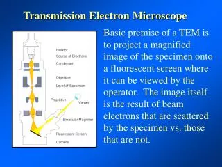
Transmission Electron Microscope

Transmission Electron Microscope
E N D
Presentation Transcript
Ernst RuskaGerman physicistHe was awarded the Nobel Prize for Physics in 1986 for building the first transmission electron microscope.
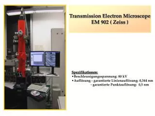
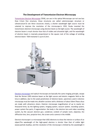
TRANSMISSION ELECTRON MICROSCOPE (TEM) • Transmission electron microscopes (TEM) are microscopes that use a particle beam of electrons to visualize specimens and generate a highly-magnified image. • Works on the same basic principle as the light microscope but uses an electron beam instead of the light beam to produce an image. • Its thickness is around 100-200nm. • Imaging in the TEM must be carried out under a vacuum, as electrons cannot travel through the air. • Using an electron microscope, we can see things that we would not normally be able to see with our naked eyes and have greater magnification than the light microscope.
COMPONENTS OF TRANSMISSION ELECTRON MICROSCOPE • Electron Gun • Vacuum System • Electromagnetic Lenses • Specimen Stage • Imaging Devices
RESOLUTION • TEMs can magnify objects up to 2 million times. • The resolution of a TEM is 1,000 times greater than a compound microscope and about 500,000 times greater than the human eye. • Transmission Electron Microscope Resolution: In a TEM, a monochromatic beam of electrons is accelerated through a potential of 40 to 100 kilovolts (kV) and passed through a strong magnetic field that acts as a lens. The resolution of a TEM is about 0.2 nanometers (nm). • The resolution of TEM is 0.5 nanometers while SEM has 0.4 nanometers.
Applications • A TEM can be used in any branch of science and technology where it is desired to study the internal structure of specimens down to the atomic level. • The TEM is used heavily in both materialscience/metallurgy and the biological sciences. • Micro Structural Analysis • Interfacial Analysis • Crystal Structure • Magnifications Up To 1,000,000 X Gt Atomic resolution • Small Region Elemental Analysis
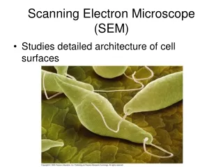
SOME IMAGES UNDER TRANSMISSION ELECTRON MICROSCOPE SEM (left) and TEM (right) images of bacteria. SEM shows numerous bacteria on a surface (green), and the TEM image shows the interior structure of a single bacterium.