Transmission Electron Microscope
50 likes | 123 Views
Creative Biogene offers an expertise transmission electron microscopy service for clients. With leading-edge transmission electron microscopes, our experienced scientist team will offer an excellent and reliable service at various stages of manufacturing process.u00a0<br>

Transmission Electron Microscope
E N D
Presentation Transcript
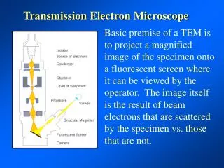
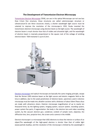
The Development of Transmission Electron Microscopy Transmission Electron Microscope (TEM), can see in the optical Microscope can not see less than 0.2um fine structure, these structures are called submicroscopic structure or ultrastructure.To see these structures clearly, a shorter wavelength light source must be selected to improve the resolution of the microscope.In 1932, Ruska invented the transmission electron microscope using electron beam as the light source. The wavelength of electron beam is much shorter than that of visible and ultraviolet light, and the wavelength of electron beam is inversely proportional to the square root of the voltage of emitting electron beam. TEM resolution is up to 0.2nm. Electron microscope and optical microscope are basically the same imaging principle, except that the former USES electron beam as the light source and electric magnetic field as the lens.In addition, due to the weak penetration of electron beam, specimens used in electron microscopy must be made into ultrathin sections with a thickness of about 50nm.These slices are made with ultramicro slicers. Electron microscope magnification of up to nearly ten thousand times, by the lighting system, imaging system, vacuum system, recording system, power system five parts, if segmentation: the body is the electron lens and video recording system, by the vacuum electron gun, condenser, content of sample room, objective, the diffraction lens, lens, projector lens, the screen and a camera in the middle. Electron microscope is a microscope that USES electrons to show the interior or surface of an object.The wavelength of the high-speed electron is shorter than that of visible light (wave-particle duality), and the resolution of the microscope is limited by the wavelength it
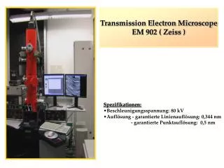
USES, so the theoretical resolution of the electron microscope (about 0.1 nm) is much higher than that of the optical microscope (about 200 nm). Transmission electron microscope (TEM), referred to as Transmission electron microscope [1], is the accelerated and concentrated electron beam projected on a very thin sample, the electron and the atom in the sample collision and change direction, resulting in solid Angle scattering.The size of the scattering Angle is related to the density and thickness of the sample, so different light and shade images can be formed, which will be displayed on the imaging devices (such as fluorescent screen, film, and photosensitive coupling components) after magnification and focusing. Because the DE Broglie wavelength of the electron is very short, the resolution of the transmission electron microscope is much higher than that of the optical microscope, which can reach 0.1 ~ 0.2nm and the magnification is tens of thousands ~ millions of times.Thus, transmission electron microscopy can be used to observe the fine structure of a sample, or even the structure of a single column of atoms, tens of thousands of times smaller than the smallest structure that can be observed with an optical microscope.TEM is an important analytical method in many scientific fields related to physics and biology, such as cancer research, virology, materials science, nanotechnology, semiconductor research and so on. At a low magnification, the contrast of TEM imaging is mainly caused by different electron absorption due to different thickness and composition of materials.When magnification power is high, the complex wave action will cause the difference in the brightness of the image, so professional knowledge is needed to analyze the obtained image.By using different modes of TEM, the sample can be imaged by means of chemical properties of the substance, crystal orientation, electron structure, electron phase shift caused by the sample, and normal electron absorption. The first TEM was developed by Max knorr and ernst lueska in 1931. The research team developed the first TEM with a resolution beyond visible light in 1933, and the first commercial TEM was successfully developed in 1939. Large transmission electron microscope Conventional TEM generally adopts 80-300kv electron beam acceleration voltage, different models correspond to different electron beam acceleration voltage, and its resolution is related to electron beam acceleration voltage, up to 0.2-0.1nm. High-end models can achieve atomic-level resolution. Low voltage transmission electron microscope Low-voltage electron microscope (LVEM) USES a much lower electron beam accelerating Voltage (5kV) than a large transmission electron microscope.The lower accelerating voltage will enhance the strength of the electron beam and the sample, thus improving the contrast and contrast of the image, especially suitable for polymer and biological samples.At the same time, the damage of low voltage transmission electron microscope to the sample is
less. The resolution is lower than large electron microscope, 1-2nm.Because of the low voltage, transmission electron microscope, scanning electron microscope and scanning transmission electron microscope can be integrated on one device Frozen electron microscopy (sem) Frozen electron microscopy (then microscopy) is usually on the ordinary transmission electron microscopy (sem) equipped with samples, refrigeration cooling to the sample of the liquid nitrogen temperature (77 k), to measure protein, biological slice is sensitive to the temperature of the samples.By freezing the sample, the damage of the electron beam to the sample can be reduced, and the deformation of the sample can be reduced, so as to obtain a more real sample morphology.