Download
1 / 59
590 likes | 795 Views
The Respiratory System. Respiration Includes. Pulmonary ventilation Air moves in and out of lungs Continuous replacement of gases in alveoli (air sacs) External respiration Gas exchange between blood and air at alveoli O2 (oxygen) in air diffuses into blood

E N D
Respiration Includes • Pulmonary ventilation • Air moves in and out of lungs • Continuous replacement of gases in alveoli (air sacs) • External respiration • Gas exchange between blood and air at alveoli • O2 (oxygen) in air diffuses into blood • CO2 (carbon dioxide) in blood diffuses into air • Transport of respiratory gases • Between the lungs and the cells of the body • Performed by the cardiovascular system • Blood is the transporting fluid • Internal respiration • Gas exchange in capillaries between blood and tissue cells • O2 in blood diffuses into tissues • CO2 waste in tissues diffuses into blood
Cellular Respiration • Oxygen (O2) is used by the cells • O2 needed in conversion of glucose to cellular energy (ATP) • All body cells • Carbon dioxide (CO2) is produced as a waste product • The body’s cells die if either the respiratory or cardiovascular system fails
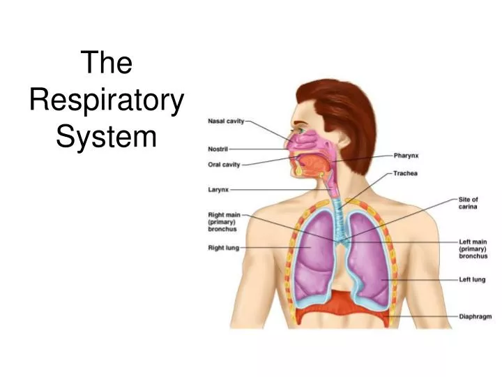
The Respiratory Organs Conducting zone • Respiratory passages that carry air to the site of gas exchange • Filters, humidifies and warms air Respiratory zone • Site of gas exchange • Composed of • Respiratory bronchioles • Alveolar ducts • Alveolar sacs Conducting zone labeled
Conducting zone will be covered first Nose • Provides airway • Moistens and warms air • Filters air • Resonating chamber for speech • Olfactory receptors External nose
Nasal cavity • Air passes through nares (nostrils) • Nasal septum divides nasal cavity in midline (to right & left halves) • Perpendicular plate of ethmoid bone, vomer and septal cartilage • Connects with pharynx posteriorly through choanae (posterior nasal apertures*) • Floor is formed by palate (roof of the mouth) • Anterior hard palate and posterior soft palate * palate
Linings of nasal cavity • Vestibule* (just above nostrils) • Lined with skin containing sebaceous and sweat glands and nose hairs • Filters large particulars (insects, lint, etc.) • The remainder of nasal cavity: 2 types of mucous membrane • Small patch of olfactory mucosa near roof (cribriform plate) • Respiratory mucosa: lines most of the cavity Olfactory mucosa *
Respiratory Mucosa • Pseudostratified ciliated columnar epithelium • Scattered goblet cells • Underlying connective tissue lamina propria • Mucous cells – secrete mucous • Serous cells – secrete watery fluid with digestive enzymes, e.g. lysozyme • Together all these produce a quart/day • Dead junk is swallowed
Nasal Conchae • Inferior to each is a meatus* • Increases turbulence of air • 3 scroll-like structures • Reclaims moisture on the way out * * Of ethmoid (its own bone) *
Paranasal sinuses • Frontal, sphenoid, ethmoid and maxillary bones • Open into nasal cavity • Lined by same mucosa as nasal cavity and perform same functions • Also lighten the skull • Can get infected: sinusitis
The Pharynx (throat) • 3 parts: naso-, oro- and laryngopharynx • Houses tonsils (they respond to inhaled antigens) • Uvula closes off nasopharynx during swallowing so food doesn’t go into nose • Epiglottis posterior to the tongue: keeps food out of airway • Oropharynx and laryngopharynx serve as common passageway for food and air • Lined with stratified squamous epithelium for protection * *
The Larynx (voicebox) • Extends from the level of the 4th to the 6th cervical vertebrae • Attaches to hyoid bone superiorly • Inferiorly is continuous with trachea (windpipe) • Three functions: • Produces vocalizations (speech) • Provides an open airway (breathing) • Switching mechanism to route air and food into proper channels • Closed during swallowing • Open during breathing
Framework of the larynx • 9 cartilages connected by membranes and ligaments • Thyroid cartilage with laryngeal prominence (Adam’s apple) anteriorly • Cricoid cartilage inferior to thyroid cartilage: the only complete ring of cartilage: signet shaped and wide posteriorly
Behind thyroid cartilage and above cricoid: 3 pairs of small cartilages • Arytenoid: anchor the vocal cords • Corniculate • Cuneiform • 9th cartilage: epiglottis
* * Posterior views Epliglottis* (the 9th cartilage) Elastic cartilage covered by mucosa On a stalk attached to thyroid cartilage Attaches to back of tongue During swallowing, larynx is pulled superiorly Epiglottis tips inferiorly to cover and seal laryngeal inlet Keeps food out of lower respiratory tract
Cough reflex: keeps all but air out of airways • Low position of larynx is required for speech (although makes choking easier) • Paired vocal ligaments: elastic fibers, the core of the true vocal cords
Pair of mucosal vocal folds (true vocal cords) over the ligaments: white because avascular
Glottis is the space between the vocal cords • Laryngeal muscles control length and size of opening by moving arytenoid cartilages • Sound is produced by the vibration of vocal cords as air is exhaled
Innervation of larynx (makes surgery at neck risky) • Recurrent laryngeal nerves of Vagus • These branch off the Vagus and make a big downward loop under vessels, then up to larynx in neck • Left loops under aortic arch • Right loops under right subclavian artery • Damage to one: hoarseness • Damage to both: can only whisper
Trachea (the windpipe) • Descends: larynx through neck into mediastinum • Divides in thorax into two main (primary) bronchi • 16-20 C-shaped rings of hyaline cartilage joined by fibroelastic connective tissue • Flexible for bending but stays open despite pressure changes during breathing
Posterior open parts of tracheal cartilage abut esophagus • Trachealis muscle can decrease diameter of trachea • Esophagus can expand when food swallowed • Food can be forcibly expelled • Wall of trachea has layers common to many tubular organs – filters, warms and moistens incoming air • Mucous membrane (pseudostratified epithelium with cilia and lamina propria with sheet of elastin) • Submucosa ( with seromucous glands) • Adventitia - connective tissue which contains the tracheal cartilages)
Carina* • Ridge on internal aspect of last tracheal cartilage • Point where trachea branches (when alive and standing is at T7) • Mucosa highly sensitive to irritants: cough reflex *
Bronchial tree bifurcation • Right main bronchus (more susceptible to aspiration) • Left main bronchus • Each main or primary bronchus runs into hilus of lung posterior to pulmonary vessels 1. Oblique fissure2. Vertebral part3. Hilum of lung4. Cardiac impression5. Diaphragmatic surface (Wikipedia)
Main=primary bronchi divide into secondary=lobar bronchi, each supplies one lobe • 3 on the right • 2 on the left • Lobar bronchi branch into tertiary = segmental bronchi • Continues dividing: about 23 times • Tubes smaller than 1 mm called bronchioles • Smallest, terminal bronchioles, are less the 0.5 mm diameter • Tissue changes as becomes smaller • Cartilage plates, not rings, then disappears • Pseudostratified columnar to simple columnar to simple cuboidal without mucus or cilia • Smooth muscle important: sympathetic relaxation (“bronchodilation”), parasympathetic constriction (“bronchoconstriction”)
Respiratory Zone • End-point of respiratory tree • Structures that contain air-exchange chambers are called alveoli • Respiratory bronchioles lead into alveolar ducts: walls consist of alveoli • Ducts lead into terminal clusters called alveolar sacs – are microscopic chambers • There are 3 million alveoli!
Gas Exchange • Air filled alveoli account for most of the lung volume • Very great area for gas exchange (1500 sq ft) • Alveolar wall • Single layer of squamous epithelial cells (type 1 cells) surrounded by basal lamina • 0.5um (15 X thinner than tissue paper) • External wall covered by cobweb of capillaries • Respiratory membrane: fusion of the basal laminas of • Alveolar wall • Capillary wall Respiratory bronchiole Alveolar duct (air on one side; blood on the other) Alveoli Alveolar sac
Bronchial “tree” and associated Pulmonary arteries
This “air-blood barrier” (the respiratory membrane) is where gas exchange occurs • Oxygen diffuses from air in alveolus (singular of alveoli) to blood in capillary • Carbon dioxide diffuses from the blood in the capillary into the air in the alveolus
Surfactant • Type II cuboidal epithelial cells are scattered in alveolar walls • Surfactant is a detergent-like substance which is secreted in fluid coating alveolar surfaces – it decreases tension • Without it the walls would stick together during exhalation • Premature babies – problem breathing is largely because lack surfactant
Microscopic detail of alveoli • Alveoli surrounded by fine elastic fibers • Alveoli interconnect via alveolar pores • Alveolar macrophages – free floating “dust cells” • Note type I and type II cells and joint membrane
Lungs and Pleura Around each lung is a flattened sac of serous membrane called pleura Parietal pleura – outer layer Visceral pleura – directly on lung Pleural cavity – slit-like potential space filled with pleural fluid • Lungs can slide but separation from pleura is resisted (like film between 2 plates of glass) • Lungs cling to thoracic wall and are forced to expand and recoil as volume of thoracic cavity changes during breathing
CXR (chest x-ray)
Chest x rays Lateral (male) Normal female
Pleura also divides thoracic cavity in three • 2 pleural, 1 mediastinal • Pathology • Pleuritis • Pleural effusion
Paired lungs occupy all thoracic cavity lateral to the mediastinum • Mediastinum contains (mainly): heart, great blood vessels, trachea, main bronchi, esophagus
Lungs • Each is cone-shaped with anterior, lateral and posterior surfaces contacting ribs • Superior tip is apex, just deep to clavicle • Concave inferior surface resting on diaphragm is the base apex apex base base
Hilus or (hilum) • Indentation on mediastinal (medial) surface • Place where blood vessels, bronchi, lymph vessel, and nerves enter and exit the lung • “Root” of the lung • Above structures attaching lung to mediastinum • Main ones: pulmonary artery and veins and main bronchus Medial view R lung Medial view of L lung
Abbreviations in medicine: e.g.” RLL pneumonia” Horizontal fissure • Right lung: 3 lobes • Upper lobe • Middle lobe • Lower lobe • Left lung: 2 lobes • Upper lobe • Lower lobe Oblique fissure Oblique fissure Each lobe is served by a lobar (secondary) bronchus
Each lobe is made up of bronchopulmonary segments separated by dense connective tissue • Each segment receives air from an individual segmental (tertiary) bronchus • Approximately 10 bronchopulmonary segments in each lung • Limit spread of infection • Can be removed more easily because only small vessels span segments • Smallest subdivision seen with the naked eye is the lobule • Hexagonal on surface, size of pencil eraser • Served by large bronchiole and its branches • Black carbon is visible on connective tissue separating individual lobules in smokers and city dwellers
Pulmonary arteries bring oxygen-poor blood to the lungs for oxygenation • They branch along with the bronchial tree • The smallest feed into the pulmonary capillary network around the alveoli • Pulmonary veins carry oxygenated blood from the alveoli of the lungs to the heart
Stroma – framework of connective tissue holding the air tubes and spaces • Many elastic fibers • Lungs light, spongy and elastic • Elasticity reduces the effort of breathing • Blood supply • Lungs get their own blood supply from bronchialarteries and veins • Innervation: pulmonary plexus on lung root contains sympathetic, parasympathetic and visceral sensory fibers to each lung • From there, they lie on bronchial tubes and blood vessels within the lungs
Understand the concepts; you don’t need to know the names of the tertiary bronchi Does this clarify a little? Primary bronchus: (Left main) • Bronchopulmonary – means both bronchial tubes and lung alveoli together • Bronchopulmonary segment – chunk receiving air from a segmental (tertiary) bronchus*: tertiary means it’s the third order in size; also, the trachea has divided three times now • “Anatomical dead space” • The conducting zone which doesn’t participate in gas exchange Secondary: (left lower lobar bronchus) (supplying left lower lobe) *
Ventilation • Breathing = “pulmonary ventilation” • Pulmonary means related to the lungs • Two phases • Inspiration (inhalation) – air in • Expiration (exhalation) – air out • Mechanical forces cause the movement of air • Gases always flow from higher pressure to lower • For air to enter the thorax, the pressure of the air in it has to be lower than atmospheric pressure • Making the volume of the thorax larger means the air inside it is under less pressure (the air has more space for as many gas particles, therefore it is under less pressure) • The diaphragm and intercostal muscles accomplish this
Muscles of Inspiration • During inspiration, the dome shaped diaphragm flattens as it contracts • This increases the height of the thoracic cavity • The external intercostalmuscles contract to raise the ribs • This increases the circumference of the thoracic cavity Together:
Inspiration continued • Intercostals keep the thorax stiff so sides don’t collapse in with change of diaphragm • During deep or forced inspiration, additional muscles are recruited: • Scalenes • Sternocleidomastoid • Pectoralis minor • Quadratus lumborum on 12th rib • Erector spinae (some of these “accessory muscles” of ventilation are visible to an observer; it usually tells you that there is respiratory distress – working hard to breathe)


