Download
1 / 29
300 likes | 1.74k Views
Aortic Stenosis & Non-Cardiac Surgery Stephen R. Ellis, MD David Warters, MD ABG: 7.58/32/472/30/8.2 Na/K 139/3.8 HCT 44 Introduction

E N D
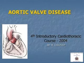
Aortic Stenosis & Non-Cardiac Surgery Stephen R. Ellis, MD David Warters, MD
ABG: 7.58/32/472/30/8.2 Na/K 139/3.8 HCT 44
Introduction • Aortic stenosis derives its position as the most important valvular lesion because of its potential for sudden death (15–20%), and because of the inability to obtain adequate systemic perfusion by external cardiac massage during a cardiac arrest.
Introduction • Aorticstenosiswithout accompanying mitral valve disease is more common in men than in women and very rarely occurs on a rheumatic basis. Instead, isolated AS is usually either congenital or degenerative in origin. • The natural history of the disease is of a long asymptomatic latent period followed by the onset of characteristic symptoms (angina, syncope, dyspnea).
Etiology • Degenerative calcific aortic stenosis • Mechanical stress over time leads to progressive fibrosis and calcification of a previously normal tri-leaflet valve. • Initially, this process is seen as sclerosis. • It is an early form of the disease that can progress to stenosis. • associated with many of the risk factors for coronary artery disease - diabetes, hypercholesterolaemia, smoking and hypertension.
Etiology • Congenital bicuspid aortic valve • Bicuspid aortic valve is the most common congenital cardiac malformation ( 2% of general population). • abnormal valve structure - two rather than three leaflets - leads to turbulent flow, which, in turn, can produce fibrosis, calcification and orifice narrowing secondary to trauma. • commonly produces symptoms in the fourth to sixth decades of life. • accounts for 50% of patients <70 yr requiring aortic valve surgery for stenosis but only 25% of those >70 yr.
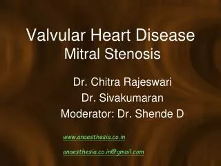
Etiology • Rheumatic AS • results from adhesions and fusions of the commissures and cusps • There is vascularization of the leaflets of the valve ring • This leads to retraction and stiffening of the free borders of the cusps. • Calcific nodules develop on both surfaces, and the orifice is reduced to a small round or triangular opening. • As a consequence, the rheumatic valve is often regurgitant and stenotic
Normal aortic valve. Congenital aortic stenosis. Rheumatic aortic stenosis. Calcific aortic stenosis. Calcific senile aortic stenosis. Etiology
Classification • Mild AS – AVA 1.2-1.8cm2, mean gradient 12-25 mmHg • Moderate AS – AVA 0.8-1.2cm2, mean gradient 40-50 mmHg • Severe AS – AVA <0.8cm2, mean gradient > 50 mmHg
Pathophysiology • The normal aortic valve area (AVA) is 2.6–3.5 cm2 in adults. • Hemodynamically significant obstruction occurs as the AVA approaches 1.0 cm2. • Increasing obstruction hypertrophy, which allows the LV to maintain a pressure gradient across the valve without dilating or reducing the cardiac output.
Pathophysiology • However, over time the hypertrophied ventricle becomes increasingly stiff, diastolic dysfunction with a reduced compliance. • This is transmitted to the pulmonary circulation pulmonary edema
Pathophysiology • A normal sinus rhythm is beneficial as the left atrial kick accounts for 40% of LV filling. • LA hypertrophies secondary to this increased demand on it increased chance of atrial fibrillation. • Major alterations of myocardial oxygen supply and demand occur. • The ventricle becomes • very sensitive to changes in preload • dependent on the maintenance of sinus rhythm • susceptible to ischemia, especially when arterial pressure is reduced.
Pathophysiology • Eventually, cardiac output, stroke volume and therefore pressure gradient across the valve fall. • Left ventricular dilatation occurs late in the disease process
There is a direct relationship b/w the aortic valve area and the flow across the valve. Blood flow is not significantly impeded until the aortic valve area is < 0.5-0.7 cm2 Pathophysiology
Assessment - Symptoms • As stated the three cardinal signs of AS are – angina, syncope, and dyspnea. • Angina • occurs as oxygen demand from the hypertrophied LV outstrips the supply • Initial symptom in 50-70% of pts • Syncope • etiology unclear • Initial symptom in 15-30% of pts
Assessment - Symptoms • Dyspnea – pulmonary congestion, CHF • Symptoms that develop late in AS, and reflect inc pulmonary HTN – exertional dyspnea, orthopnea, PND, pulmonary edema
Assessment - Exam • Arterial pulse is slow rising, and of low volume • Carotid thrill • Precordial thrill with leaning forward during expiration • Late systolic murmur (2nd intercostal space at base of heart)
Assessment - Exam • EKG: • LVH – present in ~ 85% of pts • T-wave inversion & ST depression as hypertrophy becomes worse • AV and intraventricular blocks can be seen
Assessment - Exam • ECHO: • Used to assess the anatomy of the aortic valve, grade the stenosis, and assess LV function.
Assessment - Exam • ECHO:
Anesthetic Management for Non-Cardiac Surgery • Careful hemodynamic monitoring is essential: • Arterial line • Aorticstenosis produces a fixed obstruction to left ventricular ejection that results in reduced stroke volume and an arterial pressure waveform that rises slowly (pulsus tardus) and peaks late in systole • Pulsus parvus (narrow pulse pressure) • CVC, or large bore PIVs • Swan-Ganz? Absolutely not, as the potential for it to precipitate arrhythmias is too high. • TEE is appropriate if available
Anesthetic Management for AS & Non-Cardiac Surgery • Avoid systemic hypotension • leads to myocardial ischemia, and then decreased contractility and a vicious cycle ensues. • vasoconstrictors must be at hand – consider an infusion from the beginning • treat hypotension aggressively • Maintain sinus rhythm • sinus tachy decreases diastolic time for myocardial perfusion • sinus brady limits CO in pts with fixed stroke volume
Anesthetic Management for AS & Non-Cardiac Surgery • Treat arrhythmias promptly • Contractility • Stroke volume is maintained with a heightened contractile state • Maintain adequate intravascular volume to ensure ventricular filling • b/c of dec LV compliance and inc LVEDP & LVEDV, preload augmentation is needed for a normal stroke volume
Anesthetic Management for AS & Non-Cardiac Surgery • GA vs Regional: • successful use of spinal and epidural have been reported. • Can use combined lumbar plexus and sciatic PNB for hips • GA is safe, as long as care is taken to maintain blood pressure and sinus rhythm • Narcotic-based technique is often used
Postoperative Management • Monitored bed with invasive monitoring, and adequate pain control. • Maintain appropriate intravascular filling, blood pressure and sinus rhythm.
References • Brown , J et al. Aortic Stenosis and Non-Cardiac Surgery. Continuing Education in Anaesthesia, Critical Care & Pain 2005 5(1):1-4 • Hensley F, Martin DE, Gravlee GP. A Practical Approach to Cardiac Anesthesia, 3rd ed. Philadelphia: LWW, 2003:303-309 • Miller, RD. Anesthesia, 5th ed. Philadelphia: Churchill Livingstone, 2000: 1770-1771 • Miller, RD. Anesthesia, 6th ed. Philadelphia: Churchill Livingstone, 2000: 1954-1957 • Braunwald. Heart Disease: A Textbook of Cardiovascular Medicine, 6th ed. Philadelphia: WB Saunders, 2001:1671-1680