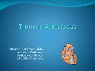
Truncus Arteriosus
Truncus Arteriosus. Seoul National University Hospital Department of Thoracic & Cardiovascular Surgery. Truncus Arteriosus. 1. Definition Congenital cardiac malformation in which one great artery, arising from the base of heart by way of a single semilunar(truncal) valve,

Truncus Arteriosus
E N D
Presentation Transcript
Truncus Arteriosus Seoul National University Hospital Department of Thoracic & Cardiovascular Surgery
Truncus Arteriosus • 1. Definition • Congenital cardiac malformation in which one great artery, arising • from the base of heart by way of a single semilunar(truncal) valve, • gives originof coronary, systemic , and one or two pulmonary • arteries proximal to the origin of the brachiocephalic branches. • Beneath the truncal valve, there is a VSD. • 2. History • Wilson : 1st description in 1798 • Buchanan : Clinical & autopsy report in 1864 • Collett & Edwards : Classification in 1949 • Van Praagh : Alternative classification in 1965 • McGoon : 1st repair with homograft in 1967
Truncus Arteriosus • Pathophysiology • A single common artery, or truncus , overlying the ventricular septum and a nonrestrictive VSD gives rise to the coronary arteries, pulmonary arteries, and ascending aorta. • Complete mixing of systemic and pulmonary venous return at the VSD and truncal valve level results in moderate cyanosis. • As the pulmonary vascular resistance decreases after birth, significant left-to-right shunting at the truncal valve level leads to excessive pulmonary blood flow, pulmonary hypertension, and congestive heart failure.
Truncus Arteriosus • Morphogenesis • Truncus arteriosus is called also as persistent truncus arteriosus, truncus arteriosus communis, common aorticopulmonary trunk • Chromosomal 22q11 deletion is present in a substantial number of patients with conotruncal abnormalities ( about one third with truncus arteriosus) • Many of these have additional characteristic features of DiGeorge syndrome, velocardiofacial syndrome, or conotruncal face syndrome • As such, their natural history may be complicated by hypocalcemia, palatal abnormalities, learning disability, or other noncardiac problems.
1. Truncal artery 2. Pulmonary arteries Type I, II : 80~90% III, IV : 5~10% Hemitruncus : 2% Stenosis of origin : 10% 3. Aorta & ductus arteriosus Wide PDA : Arch hypoplasia, IAA or CoA(10~15%) No PDA : majority 4. Coronary arteries 5. Semilunar valve Tricuspid (1/2~2/3), quadricuspid, bicuspid (5%) Myxomatous thickening (1/3) Truncal stenosis (20%) 6. VSD (juxtatruncal) 7. RV (absent conal septum) 8. Left ventricle ; normal 9. Associated anomalies IAA or CoA with PDA : 10~15% RAA : 25~35% Anomalous branch : 10% LSVC : 10% ASD : 10% DiGeorge synd. AV discordance, situs inversus, heterotaxia, DILV, MS, AV-canal, tricuspid stenosis Morphology of Truncus Arteriosus
Classification of Truncus Arteriosus (Collett & Edwards)
Truncus Arteriosus Truncal valve VSD
Clinical Features & Diagnosis • 1. Symptoms • tachypnea, tachycardia, irritability, mild cyanosis • 2. Physical examination • signs of CHF, overactive heart • truncal insufficiency (systolic & diastolic murmur) • stenosis of PA (continuous murmur) • 3. Chest radiography • marked cardiomegaly as well as plethora • 4. EKG • RAD, biventricular hypertrophy • 5. Echocardiography • 6. Cardiac catheterization & cineangiography
Truncus Arteriosus • Natural History • 1. Incidence • rare, unfavorable natural history • 1.7% to 2.8% of CHD • 2. Survival • 50% survival in 1 month • 18% survival in 6 months • 12% survival in 1 year • Others : Eisenmenger syndrome (death in 3rd decade) • 3. Modes of death • . Congestive heart failure in early life • . SBE, cerebral abscess, pulmonary vascular disease • . Survival is favorably affected by PS
Operative Techniques • 1. Repair with allograft valved conduit • 2. Repair truncus I,II with autologous tissue • Barbero-Marcial technique • 3. Repair of hemitruncus • Unifocalization of pulmonary artery • 4. Repair of truncus arteriosus with IAA
Operative Indications • 1. Diagnosis of truncus is an indication for it’s repair; • because about 50% of surgically untreated patients • die in the 1st month of life. • 2. Repair should be recommended as early in life • as possible rather than deferring to some • predetermined age • 3. Importantly elevated PVR is a contraindication • in old infant (more than 6~12 months old).
Truncal Valve Remodeling Technique • Diagrams shows truncal valve repair by leaflet excision • and annular remodeling, usually there is one leaflet • that is grossly prolapsed
Surgical Results of Truncus Arteriosus • 1. Survival • Early death • Time-related survival • 2. Modes of death • 3. Incremental risk factors for premature death • 1) Age at repair 2) Functional class • 3) Type 4) Size of VSD • 5) Predominance of origin of truncal artery • 6) Small size of pulmonary arteries • 7) Truncal valve abnormalities • 8) Hemitruncus • 9) Major associated cardiac anomalies • 10) Pulmonary vascular disease • 4. Progressing truncal valve incompetence • 5. Conduit reoperation