Download
1 / 12
280 likes | 2.89k Views
Kommerell’s Diverticulum Repair with a Right-Sided Aortic Arch: Strategies and Techniques. Phillip D. Smith, MD Cardiac Surgery Research Fellow Cardiothoracic Surgery University of Colorado Wilson Y. Szeto, MD Assistant Professor, Cardiovascular Surgery University of Pennsylvania

E N D
Kommerell’s Diverticulum Repair with a Right-Sided Aortic Arch: Strategies and Techniques Phillip D. Smith, MD Cardiac Surgery Research Fellow Cardiothoracic Surgery University of Colorado Wilson Y. Szeto, MD Assistant Professor, Cardiovascular Surgery University of Pennsylvania T. Brett Reece, MD Assistant Professor, Cardiothoracic Surgery University of Colorado
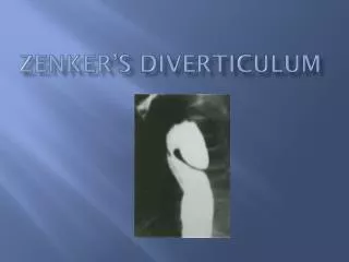
Background • Kommerell’s Diverticulum • Congenital diverticulum of a subclavian artery associated with various aortic pathologies and aberrant subclavian anatomy • Right sided Aortic Arch • Two types • Felson and Palayew Type 1- mirror image anatomy of the great vessels • Felson and Palayew Type 2- aberrant left subclavian Backer, CL. Eur J Cardiothorac Surg 2002;22:64-69
Case Report • Three patients with a right-sided aortic arch and Kommerell’s diverticulum of an aberrant left subclavian artery • All greater than 4 cm in maximum diameter • Two males in their fifties presenting with dysphagia • One 36 year old female presenting with dyspnea secondary to tracheal compression • All healthy, no significant past medical history
Representative 3D CTA showing Kommerell’s diverticulum and aberrant left subclavian artery Sagittal view showing compression of the trachea by the arch and aberrant subclavian artery
Goals of Operation • Eliminate risk of rupture and/or dissection • Relief of symptoms, while desired, should not be expected • Patients should understand that symptoms may not be ameliorated after procedure • May be accomplished with resection or exclusion of the diverticulum
Potential Operative Approaches • TEVAR exclusion • Revascularization of the aberrant subclavian artery • Isolation of diverticulum from blood flow • Left Thoractomy • Easier to address distal aberrant subclavian artery • Descending aortic exposure difficult • Right Thoracotomy • Subclavian artery revascularization +/- ligation of proximal subclavian artery • Most direct approach to right sided descending aorta • Left Heart Bypass versus Full Heart Bypass versus HCA • Depends on exposure for arch cross clamp
Our Operative Approach • Two Stage Procedure • I: Subclavian revascularization via proximal ligation and transposition • II: Descending thoracic aortic reconstruction • Using CPB or HCA • Diverticulum resection
Reasoning for this Technique • Definitive treatment of diverticulum by resection • Inadequate landing zone in these cases due to proximity of brachiocephalic vessels and extreme angulation of distal arch • Potential use of HCA as aortic tissue around the diverticulum is extremely friable
Outcomes • Postoperative CT reconstruction showing resected diverticula and flow to the left subclavian via the transposition graft • Both cases of dysphagia were completely resolved at one year • Tracheal compression much improved in dyspneic patient with improved DOE
Left: Sagittal CT showing resolution of tracheal compresion Top Right: Posterior 3D CTA reconstruction showing resected diverticulum and subclavian transposition with flow Bottom Right: Anterior 3D CTA showing flow in left subclavian after transposition
Key Points Goals of treatment are prevention of aortic catastrophe, not treatment of symptoms While minimally invasive approaches are feasible, this series demonstrates that a staged open approach is safe and may provide symptomatic relief in addition to aortic protection
Conclusion A staged open approach to Kommerell’s Diverticulum resection can be safely performed with minimal morbidity and potential symptomatic relief