Download
1 / 89
940 likes | 1.58k Views
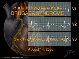
Sudden Cardiac Arrest BRUGADA SYNDROME. Carlo Francisco S. Gochuico, M.D. August 14, 2008. OBJECTIVES. To present a case of sudden cardiac arrest in a young male To discuss the approach and management of Brugada syndrome. General Data. J.D. 28 year old Male Filipino Chief Complaint

E N D
Sudden Cardiac Arrest BRUGADA SYNDROME Carlo Francisco S. Gochuico, M.D. August 14, 2008
OBJECTIVES • To present a case of sudden cardiac arrest in a young male • To discuss the approach and management of Brugada syndrome
General Data • J.D. • 28 year old • Male • Filipino Chief Complaint Loss of consciousness
One hour PTA Found unconscious Seen with upward rolling of eyeballs, stiffening of extremities, and salivary pooling Lasted 3 to 5 minutes Regained full consciousness with no recollection of the incident Was able to drink, sit on a chair, and talk with parents No slurring of speech, no extremity weakness, no chest pain History of Present Illness
Five minutes after Recurrence of stiffening of extremities and upward rolling of eyeballs Rushed to MMC ER During transit, he remained unconscious History of Present Illness
Events at ER • Quicklook showed cyanosis • BP 0 HR 0 • GCS 3 E1V1M1 • Initial tracing at ER at 1:42 AM • Chest compressions and bag mask ventilation • Defibrillation at 200 joules • Epinephrine 1mg IV • Intubated and advised ICU admission • CXRAY, stat5 and ABG
12 L EKG 8-1-08 2:16 AM post defibrillation CRBBB PR interval 0.16 sec QT interval 0.36 sec
Events at ER • 2:33 AM at ER • Systolic BP170 • Pulse 80s regular • Post CP arrest
Events at ER • 2:47 AM • Episode of stiffening of extremities • chest compression and defibrillation at 200 joules
Events at ER • 2:52 AM • Post defibrillation • BP 120/80 • Pulse 80s • Amiodarone drip started • Cardiac enzymes • Referred to neurology • Admitted to ICU
Review of Systems • General: no fever, fatigue, weight loss • Skin: no rashes, jaundice, ecchymoses, petechiae • Head: no recent head injury, headache, dizziness • Eyes: no blurring of vision, redness, pain • Ears: no tinnitus, vertigo, earache, discharge
Review of Systems • Nose: no colds, nasal congestion, discharge • Mouth: no sore throat, hoarseness • Neck: no pain, lumps, mass, stiffness • Respiratory: no cough, dyspnea, wheezing, hemoptysis • Cardiac: no chest pain, palpitations, orthopnea, PND, edema, recent chest trauma
Review of Systems • Gastrointestinal: no nausea, vomiting, regurgitation, dysphagia, hematemesis, abdominal pain, change in frequency and characteristic of stools • Urinary: no hematuria, dysuria, oliguria, polyuria, urgency, hesitancy • Vascular: no claudication, varicose veins, leg cramps • Musculoskeletal: no myalgia, arthralgia, and swelling
Review of Systems • Hematologic: no anemia, pallor, easy bruising or bleeding • Endocrine: no heat or cold intolerance, no excessive thirst or hunger • Psychiatric: no depression, nervousness
Past Medical History • No history of HPN, DM, and BA • PTB, treated for 6 months • No previous seizure disorder, no known neurologic and cardiac problems • Had a history of syncope during early childhood and another one a year ago, no work-ups were done
Family History • (+) HPN in paternal side • (-) DM, BA, stroke, cancer • No seizure disorder
Personal and Social History • Occasional smoker • Occasional alcoholic beverage drinker • Denies illicit drug use
PHYSICAL EXAMINATION • Conscious, restless, intubated • BP 120/80 HR 80 regular RR 20 assisted afebrile • Warm moist skin, no active dermatoses, no jaundice • Anicteric sclerae, pink palpebral conjunctivae, pupils 2-3 mm ERTL OU • No visible anterior neck mass, no neck vein distention, no carotid bruit • No cervical lymphadenopathies
PHYSICAL EXAMINATION • Equal chest expansion, no retractions, no rales, wheezes, crackles • Adynamic precordium, AB at 5th LICS MCL, regular rhythm, S1>S2 at base, S2>S1 at apex, no extra heart sounds, no murmurs • Abdomen flabby, normoactive bowel sounds, soft, no guarding, no direct and rebound tenderness, no hepatosplenomegaly • No costovertebral angle tenderness • No pedal edema • Pulses full and equal
PHYSICAL EXAMINATION • GCS 11 E4V1 (intubated) M6 • No papilledema • Full and equal EOM • No facial asymmetry • Intact doll’s eye movement, gag and corneal reflexes • Direct tendon reflexes normal • Motor and Sensory: intact • Localizes to pain and temperature • No babinski, kernig’s, and brudzinski
28 M Filipino Loss of consciouness at night during sleep 2 episodes of stiffening of extremities Post CP arrest 2x (VFib) No fever, headache, chest pain, weakness History of syncope 2x Positive family history of cardiac disease No illicit drug use Salient Features
Differential Diagnosis • Structural heart diseases • Ischemic heart disease • Non-ischemic cardiomyopathies • Valvular heart diseases • Arrhythmogenic right ventricular dysplasia • Primary electrophysiologic abnormalities • Long QT syndrome • Brugada syndrome
Ischemia • Cardiac arrest due to ventricular arrhythmias may be due to chronic scar or to acute MI/ischemia. A chronic infarct scar can serve as the focus for reentrant ventricular tachyarrhythmias
Non-ischemic cardiomyopathies • represent the second largest group of patients who experience SCD • Dilated cardiomyopathy is usually characterized by ventricular dilatation, initially usually of the left ventricle (LV), with myocyte hypertrophy and diminished systolic function
Hypertrophic cardiomyopathy (HCM) • an autosomal-dominant, incompletely penetrant genetic disorder resulting from a mutation in one of the many (>45) genes encoding proteins of the cardiac muscle sarcomere
Echo of HCM • Small LV cavity due to marked hypertrophy of the myocardium and encroachment into the LV cavity • Reduced septal motion and thickening during systole, particularly of the upper septum, due to the disarray of the myofibrillar architecture and abnormal contractile function • Reduced rate of closure of the mitral valve in mid diastole due to a decrease in LV compliance Left atrial enlargement
Course in the Ward • 1st Hospital day in the ICU (1230 PM) • Repeat 12L EKG done • Rsr’ pattern • ST elevation in lead V1-V3 • 2D echo – N LVD EF69% Normal LA and RA dimensions • Normal MV, TV, AV, PV • Mild MR, TR • Normal left ventricular diastolic function indices • FIo2 adjusted to 0.35
Course in the Ward • ICU at 1500H • T-piece placed and repeat ABG done • Referred to cardiology EPS • Thyroid function test requested • Quinidine bisulfate started
Course in the Ward • ICU at 1750 H • Patient extubated • 4th Hospital day • Transferred to regular room • Quinidine 200mg/tab adjusted to 1 ½ tablets in the morning and evening, and 1 tablet at lunchtime
Course in the Ward • 5th hospital day • Repeat 12 L EKG • Normal • Discharged on 6th hospital day • Follow-up after 1 week
CXRAY Aug 1, 2008 • Lungs are clear • Heart is magnified • ET in place with tip at carina
Cranial CT Scan Aug 1, 08 • Normal non-contrast CT of the brain • EEG • Normal
2D ECHOCARDIOGRAPHYAugust 1, 2008 • Normal left ventricular dimension with normal wall motion and contractility. • Computed LV EF 69% • Normal left and right atrial dimensions • Normal mitral, aortic, tricuspid, and pulmonic valves • Mild MR, TR • Normal left ventricular diastolic function indices
Repeat 12 L EKG 8-1-08 12:30 PM IRBBBPR interval 0.16 secQT interval 0.40 sec
CBC Hgb 16.8 Hct 50.2 WBC 14.21 seg54 lymph36 eos4 mono5 Baso1 Platelet 204000 Stat 5 Na 137 K 2.7 Hgb 16.7 Hct 49 Glucose (random) 245 mg/dL Laboratory Results and Ancillary Procedures
PTT patient 27.3 control 27.6 PT patient 11.1 control 11.7 activity 115.6% INR 0.94 Laboratory Results and Ancillary Procedures Cardiac enzymes CK total 165 U/L CPK MB 1.4 ng/mL Troponin I 0 ng/mL
glucose 120 CPK 506 LDH 262 ALT 185 AST 82 alkaline phosphatase 148 SPEC 23 Na 137 K 3.6 Cl 103 calcium 9.78 BUN 13 creatinine 1.1 HDL 46 trig 119 LDL 166 cholesterol 245 Laboratory Results and Ancillary Procedures
Routine urinalysis Yellow hazy PH 6 spgr 1.020 +3 protein trace sugar negative ketones, nitrites, leucocyte esterase +1 blood rbc 2/hpf wbc 2/hpf epith 10/hpf Bact 15/hpf Thyroid Function Test TSH0.037(0.27-3.75) FT34.419 (4.2-12) FT417.815 (8.8-33) Laboratory Results and Ancillary Procedures
ABG Post intubation PO2 426.2 PH 7.34 PCO2 41 HCO3 22.1 O2 sat 99.8 AC mode 100% FiO2 VT 420 Laboratory Results and Ancillary Procedures
Laboratory Results and Ancillary Procedures • ABG • 5 PM • PO2 169.1 • PH 7.39 • PCO2 37.3 • HCO3 22.5 • O2 sat 99.1 • inline neb via T piece 35% FiO2
Repeat 12 L EKG 8-6-08 9:54 AM NormalPR interval 0.20 secQT interval 0.44 sec
DISCUSSION • Sudden cardiac death (SCD) is an unexpected death due to cardiac causes, heralded by loss of consciousness occurring in a short time period (generally within 1 hour of symptom onset) • Most cases are due to cardiac arrhythmias such as VF or VT which is responsible for 50-80% of cases
Sudden Cardiac Death • Most cases of SCD occur in patients with structural abnormalities in the heart, related to either a prior MI, coronary artery disease, cardiomyopathies • Valvular diseases such as aortic stenosis are associated with increased risk of SCD • Acute inflammatory and infiltrative disorders, such as myocarditis, provide a sustained risk of SCD
Sudden Cardiac Death • Less commonly, SCD happens in patients who may not have apparent structural disease • These conditions are usually inherited arrhythmia syndromes or primary electrophysiologic abnormalities
Primary Electrophysiologic Abnormalities • This generally represents a group of abnormalities in which patients have no apparent structural heart disease but have a primary electrophysiologic abnormality that predisposes them to VF or VT • Brugada syndrome, Long QT syndrome