Download
1 / 56
560 likes | 643 Views
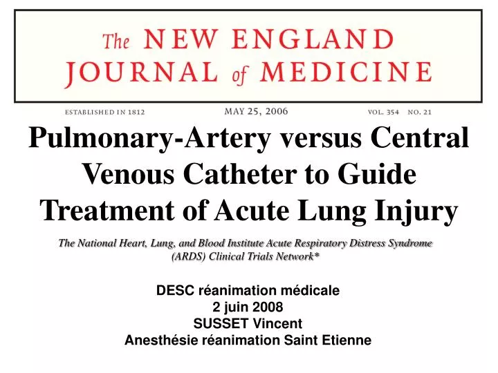
Pulmonary-Artery versus Central Venous Catheter to Guide Treatment of Acute Lung Injury. The National Heart, Lung, and Blood Institute Acute Respiratory Distress Syndrome (ARDS) Clinical Trials Network*. DESC réanimation médicale 2 juin 2008 SUSSET Vincent

E N D
Pulmonary-Artery versus Central Venous Catheter to Guide Treatment of Acute Lung Injury The National Heart, Lung, and Blood Institute Acute Respiratory Distress Syndrome (ARDS) Clinical Trials Network* DESC réanimation médicale 2 juin 2008 SUSSET Vincent Anesthésie réanimation Saint Etienne
Comparaison du monitorage hémodynamique par cathétérisme cardiaque droit (PAC) ou par mesure de PVC chez les patients en acute lung injury et SDRA Comparaison de 2 régimes de remplissage chez les patients en acute lung injury et SDRA Essai multicentrique controlé comparatif doublement randomisé en ouvert PAC 1° randomisation informatisée par bloc de 8 PVC conservative use of fluids 2° randomisation liberal use of fluids stratifiée par centre et par affectation à un système de monitorage
2 objectifs • critère primaire: différence de mortalité à 60 j • critères secondaires: temps passé sous assistance respiratoire, morbidité, critères intermédiaires (PAM, recours aux vasopresseurs…) • par utilisation du PAC vs PVC • 2) par utilisation d’un protocole de remplissage
Critères d’inclusion PaO2 / FiO2 < 300 Opacités pulmonaires bilatérales / RP Recours à la ventilation mécanique Exclusion clinique des défaillances gauches
Critères d’exclusion principaux Age < 13 ans IDM < 30j Présence du PAC avant l’inclusion ALI non inclu dans les 48h suivant le diagnostic Impossibilité d’obtenir un consentement Atteinte organique chronique (rénale, neuromusculaire, pulmonaire…) associée à une mortalité > 50% dans les 6 mois
20 centres Juin 2000 à oct 2005
Non précisé: la proportion dans chaque groupe du régime de remplissage attribué par la deuxième randomisation!!!
Mortalité à 60j identique: PAC 27,4% vs PVC 26,3% p = 0,69 Nombre de jours sans assistance respiratoire à 28j: PAC 13.2±0.5 vs PVC 13.5±0.5 p = 0.58
Suivi du protocole similaire dans chaque groupe PAC et PVC: 91±1% et 88±1% respectivement P = 0.12 Utilisation de dobutamine significativement différente PAC 7% PVC 2% P<0.001) Pas de différence concernant les paramètres ventilatoires sur les 7 premiers jours Pas de différence sur l’utilisation de cristalloïdes, colloïdes et GR ( données non fournies) Pas de différence sur le recours à l’épuration extra rénale
brièvement Résultats De la deuxième partie de l’étude: Morbi-mortalité en fonction du régime de remplissage
Mortalité à 60j identique: 25,5% conservativ gr vs 28,4% liberal gr, p=0,3 Nombre de jours sans assistance respiratoire à 28j: 14.6 conservativ gr vs 12.1 liberal gr, P < 0.001 Études chez l’homme et l’animal: attitude de restriction hydrique dans l’ARDS diminue le temps passé en réanimation et améliore les gaz du sang Ali J, et al. J Clin Invest. 1979 ; Long R, et al. 1988 ; Mitchell JP, et al Am Rev Respir Dis. 1992 ; Martin GS, et al 2002 ; Schuller D, et al 1991
Validité interne: réalité statistique du résultat Mortalité à 60j identique: PAC 27,4% vs PVC 26,3% p 0,69 Nombre de jours sans assistance respiratoire à 28j: PAC 13.2±0.5 vs PVC 13.5±0.5 p 0.58 Risque α fixé à 5% Comparaison données de survie / z-test Résultat non issu d’une analyse en sous groupe Calcul au préalable de l’effectif nécessaire portant sur le critère primaire Pas de définition à posteriori de l’hypothèse testée
Validité interne: recherche des biais Biais de confusion: a priori NON; essai comparatif, le groupe PVC sert de gr control et utilisation d’un protocole de ventilation protectrice identique dans chaque groupe Biais de sélection: POSSIBLE; malgré double randomisation, plus de patients choqués et sous vasopresseurs dans le groupe PAC et délai à l’instauration du protocole de remplissage plus important Rq: nombre de patients alloués dans chaque protocole de remplissage non précisé (même si stratification par bloc de 8…) Biais de suivi: OUI; étude en ouvert
Validité interne: recherche des biais Biais de confusion: a priori NON; essai comparatif, le groupe PVC sert de gr control et utilisation d’un protocole de ventilation protectrice identique dans chaque groupe Biais de sélection: POSSIBLE; malgré double randomisation, plus de patients choqués et sous vasopresseurs dans le groupe PAC et délai à l’instauration du protocole de remplissage plus important Rq: nombre de patients alloués dans chaque protocole de remplissage non précisé (même si stratification par bloc de 8…) Biais de suivi: OUI; étude en ouvert
Validité interne: recherche des biais Biais d’évaluation: NON; pour le critère principal: car mortalité = critère objectif POSSIBLE pour les critères secondaires dont certains sont subjectifs, sevrabilité de l’assistance respiratoire… Biais d’attrition: LIMITES; car pas de perdu de vue, peu d’inobservance de protocole ou de changements de groupe, et analyse en intention de traiter
Cohérence externe Méta-analyses et utilisation du PAC Pulmonary artery catheters for adult patients in intensive care. Pas d’efficacité démontrée, 1 seule étude identifiée ayant une puissance suffisante Harvey S et al; Cochrane Database Syst Rev. 2006 Routine perioperative pulmonary artery catheterization has no effect on rate of complications in vascular surgery: a meta-analysis. Résultats des études homogènes, pas de gain sur la morbi-mortalité Barone JE et al;Am Surg 2001 The incidence of major morbidity in critically ill patients managed with pulmonary artery catheters: a meta-analysis.Amélioration de la morbidité chez les patients monitorés avec PAC Ivanov R et al; Crit Care Med. 2000 Impact of the pulmonary artery catheter in critically ill patients: meta-analysis of randomized clinical trials. Pas de protocole défini permettant de montrer un bénéfice avec l’utilisation du PAC Shah MR et al;JAMA. 2005
Cohérence externe Harvey S et al; Cochrane Database Syst Rev. 2006
Cohérence externe Harvey S et al; Cochrane Database Syst Rev. 2006
Cohérence externe Harvey S et al; Cochrane Database Syst Rev. 2006 Harvey S et al; Cochrane Database Syst Rev. 2006
Pertinence clinique Critère principal: mortalité à 60 j, bien défini, non composite, pertinent et objectif • Intérêt de la comparaison avec la PVC? • PVC pertinente pour les valeurs < 5 mmHg; au dessus? • comparaison aux paramètres dynamiques POD ou PAPO pas plus informatifqu'un simple tirage au sort pour prédire la réponse à une épreuve de remplissage Michard. Am J Respir CCM 2000
Pertinence clinique Reproductibilité des protocoles de monitorage et de remplissage possible puisque bien détaillés dans l’appendice de l’étude Protocoles simples basés sur critères cliniques et hémodynamiques Population de l’essai: Critères d’inclusion larges: ALI ou SDRA
Pertinence clinique Population de l’essai: Cr exclusion nombreux en particuliers les patients transplantés (chimiothérapie de conditionnement…) > 10000 patients exclus!! → pas complètement représentative?? →20% des patients exclus parce que déjà monitorés par PAC avant leur inclusion dans l’étude: les patients les plus graves ou peut-être ceux à qui le PAC bénéficierait le plus ont été exclus? Biais de sélection? Résultats non extrapolables pour des patients présentant d’autres pathologies: régimes de pression intra thoracique et transmurale profondément modifiés chez les patients en SDRA
Pertinence clinique et conclusion Balance bénéfice-risque en défaveur du PAC Le cathétérisme cardiaque droit, ne doit pas être proposer à tous les patients en SDRA puisque la mortalité n’est pas améliorée en revanche la morbidité imputable à la technique est multipliée par 2 Quels patients? Problèmes de l’interprétation des données Quel monitorage et quel protocole? Méta-analyse sur données individuelles…
4.3 Exclusion Criteria 1. Absence of current intent or ability on the part of the treating physician(s) to obtain or continue central venous access (that could be used for either CVC or PAC monitoring) as part of regular care of this patient. 2. Unwillingness or inability to utilize the low tidal volume (6ml/kg PBW) ventilator management protocol. 3. Presence of a PAC at any time after onset of ALI (meeting of inclusion criteria 1-4). 4. > 48 hours since onset of ALI (meeting of inclusion criteria 1-4). 5. Age < 13 years. 6. Burns > 40% body surface area. 7. Not committed to full support (Exception: a patient will not be excluded if he/she would receive all supportive care except for attempts at resuscitation from cardiac arrest). 8. Bone marrow transplantation. 9. Acute myocardial infarction within the last 30 days. 10. Severe chronic respiratory disease: • FEV1 less than 20 ml/kg PBW (e.g., 1.4 L for 70 kg), or • FEV1 /VC less than 50% predicted, or • Chronic hypercapnia (PaCO2 greater than 45 mmHg) and/or chronic hypoxemia (PaO2 < 55 mmHg) on FiO2 = 0.21, or • Radiographic evidence of chronic over-inflation or chronic interstitial infiltration, or • Hospitalization within the past six months for respiratory failure (PaCO2 > 50 mmHg or PaO2 < 55 mmHg or O2-Sat < 88% on FiO2 = .21). • Chronic restrictive, obstructive, neuromuscular, chest wall or pulmonary vascular disease resulting in severe exercise restriction, e.g., unable to climb stairs or perform household duties, secondary polycythemia, severe pulmonary hypertension (mean PAP > 40 mmHg), or respirator dependency. 11. Neuromuscular disease that impairs ability to ventilate spontaneously, such as C5 or higher spinal cord injury, amyotrophic lateral sclerosis, Guillain-Barre Syndrome, and myasthenia gravis. 12. Morbid obesity (> 1kg/cm body weight). 13. Malignancy or other irreversible disease or condition for which 6 month mortality is estimated to be 50%. 14. Vasculitis with diffuse alveolar hemorrhage. 15. Pregnancy (negative pregnancy test required for women of child-bearing potential). 16. Renal failure requiring renal replacement therapy. 17. Severe, chronic liver disease (Child-Pugh Score of 10-15 See Appendix F). 18. Furosemide allergy. 19. Lung transplantation.
D. Fluid Bolus (Cell #19 only): 1. Withhold fluid bolus if: Cardiac index (CI) is greater than or equal to 4.5 OR FiO2 is greater than or equal to 0.7. 2. Use 15 ml/kg PBW normal saline, Plasmalyte, or Ringer's lactate (rounded to the NEAREST 250 cc) or 1 unit of RBCs or 25 grams albumin (physicians discretion) over less than or equal to 1 hour then reassess patient within 4 hours. Administer up to 3 boluses over 24 hours if indicated by protocol. This 24 hour period begins with the first protocol-mandated non-shock bolus OR the first protocol- mandated bolus following shock reversal. 3. Additional fluid boluses are allowed at the discretion of the physician. E. KVO IV: 1. Also minimize as much as possible all other fluid volume (e.g., for delivery of antibiotics etc.), except as required for nutrition support. F. Guidelines for Management of Shock: § Shock is defined as a MAP < 60 mmHg or a MAP > 60 while receiving vasopressors. § Assessments during shock should be recorded at least every 4 hours and at the time of each new entry or exit from a shock cell (cells 1 and 2). § Physicians have the choice of either fluid bolus and/or vasopressor therapy (in any order) as follows: 1. Fluid Bolus (Shock): Use 15 ml/kg PBW normal saline, Plasmalyte, or Ringers (rounded to the NEAREST 250 cc) or 1 unit of RBCs or 25 grams albumin (physicians discretion) over less than or equal to 1 hour then reassess patient. 2. Vasopressor Therapy: Choice of any single agent or any combination of the following: a. Dopamine 5 mcg/kg/min, increase to a maximum of 25 mcg/kg/min. b. Norepinephrine at 1 mcg/min, increase to a maximum of 100 mcg/min c. Epinephrine at 1 mcg/min, increase to a maximum of 20 mcg/min. d. Phenylephrine at 10 mcg/min, increase to a maximum of 500 mcg/min. e. Intravenous Vasopressin 0.005-0.04 international units/minute 3. Vasopressor Weaning (includes any dose of dopamine): a. When MAP > 60 mmHg on a stable dose of vasopressor, begin reduction of the vasopressor by greater than or equal to 25% of the stabilizing dose at intervals of less than or equal to 4 hours to maintain MAP greater than or equal to 60 mmHg. b. Dopamine is considered "discontinued" for vasopressor use and cell assignment when it is weaned to less than or equal to 5 mcg/kg/min, but should continue to be weaned per protocol (footnote F.3.a. above). G. Invalid PAOP 1. If a valid PAOP measurement cannot be obtained, use the pulmonary artery diastolic pressure to estimate the PAOP, based upon the most recently available relationship between PAOP and PADP, and assuming a stable arithmetic difference between the two values. For example, if the most recent prior valid measurements showed a PAOP = 10 and a PADP = 15, and the current PADP = 20 and a valid PAOP cannot be obtained, then assume a current PAOP = 15. 2. If neither a valid PAOP nor PADP can be obtained, then utilize the current CVP value. * Renal Failure a. Dialysis dependence is defined as the period from the initiation of dialysis to the time that continuous dialysis is discontinued or to the time following the last session of intermittent dialysis. b. During this period of dialysis dependence, urine output is considered to be adequate for the purposes of cell assignment.
Statistiques Puissance de 90% pour mettre en évidence une différence de 10% sur le critère primaire, en incluant 1000 patients (calcul de l’effectif avant le début de l’étude) Analyse de survie par méthode de Kaplan–Meier z-test pour la comparaison des données de survie Analyse en intention de traiter
Supplemental Table 2: Organ Failure Free Days to Day 7 and 28 Day 7 Day 28 Organ system PAC CVC p value PAC CVC p value Cardiovascular 4.1+/-0.1 4.1+/-0.1 0.96 19.1+/-0.4 19.0+/-0.5 0.98 Central Nervous System 3.1+/-0.2 3.3+/-0.2 0.35 17.9+/-0.5 18.1+/-0.5 0.80 Coagulation 5.3+/-0.1 5.6+/-0.1 0.09 21.5+/-0.4 22.1+/-0.4 0.33 Hepatic 5.6+/-0.1 5.6+/-0.1 0.71 21.7+/-0.4 21.5+/-0.5 0.78 Renal 5.4+/-0.1 5.7+/-0.1 0.12 21.1+/-0.5 21.7+/-0.5 0.32
Appendice Deuxième partie de l’étude
Results:The mean seven-day cumulative fluid balance was –136 ± 491 mL for the conservative group and +6992 ± 502 mL for the liberal group (P < 0.001). Diuretic-use was significantly higher in the conservative group (41% compared to 10% in the liberal group (P < 0.001). Dobutamine utilization was similar in both groups (4% vs 6%). There was no significant difference in primary outcome: 60-day mortality was 25.5% in the conservative group vs 28.4% in the liberal group (P = 0.3). However, patients in the conservative group had more ventilator-free days (14.6 vs 12.1, P < 0.001), more ICU-free days (13.4 vs 11.2, P < 0.001) and fewer days with central nervous system (CNS) failure (17.2 vs 18.8, P = 0.03) over 28 days. The liberal group had more days free of cardiovascular failure (4.2 vs 3.9, P = 0.04). There was no difference in dialysis requirement. The conservative group had lower mean arterial pressure, stroke volume and cardiac index. However, there was no difference in heart rate, venous oxygen saturation or percentage of patients receiving vasopressors. While there was an increase in metabolic alkalosis and hypokalemia for the conservative group (42 vs 19 events), there was no increase in dysrhythmias. The results were also independent of allocation to CVP vs PAOP monitoring in the concurrent trial.1