Download
1 / 63
900 likes | 1.65k Views
HYDROCEPHALUS. W & W 495-503. Hydrocephalus. A syndrome, or sign, resulting from disturbances in the dynamics of cerebrospinal fluid (CSF), which may be caused by several diseases. Incidence. Occurs in 3-4 of every 1000 births. Cause may be congenital or acquired.

E N D
HYDROCEPHALUS W & W 495-503
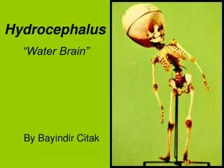
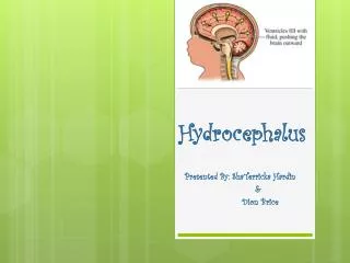
Hydrocephalus • A syndrome, or sign, resulting from disturbances in the dynamics of cerebrospinal fluid (CSF), which may be caused by several diseases.
Incidence • Occurs in 3-4 of every 1000 births. • Cause may be congenital or acquired. • Congenital- may be due to maldevelopment or intrauterine infection • Acquired- may be due to infection, neoplasm or hemorrhage.
Pathophysiology • CSF is formed by two mechanisms: • Secretion by the choroid plexus, • Lymphatic-like drainage by the extracellular fluid in brain. CSF circulates thru ventricular system and is absorbed within subarachnoid spaces by unknown mechanism.
Mechanisms of Fluid Imbalance • Hydrocephalus results from: • 1. Impaired absorption of CSF within the subarachnoid space (communicating hydrocephalus), or • 2. Obstruction to the flow of CSF through the ventricular system (non-communicating hydrocephalus)
Mechanisms of fluid imbalance • Both lead to increase accumulation of CSF in the ventricles! • Ventricles become dilated and compress the brain. • When this happens before cranial sutures are closed, skull enlarges. • In children <10-12, previously closed sutures may open.
Hydrocephalus • Most cases of non-communicating (obstructive) hydrocephalus are a result of developmental malformations. • Other causes: neoplasms, intrauterine infections, trauma. • Developmental defects account for most causes of hydrocephalus from birth to 2 years of age. (Table 11-3, page 497- sites and types of hydrocephalus)
Common Defects • Arnold-Chiari Malformation (ACM) • Type 2 malformation of brain seen most exclusively with myelomeningocele, is characterized by herniation of a small cerebellum, medulla, pons, and fourth ventricle into the cervical spinal canal through an enlarged foramen magnum.
Clinical manifestations • Clinical picture depends on acuity of onset and presence of preexisting structural lesions.
Infancy • Head grows at alarming rate with hydrocephalus. • First signs- bulging of fontanels without head enlargement. • Tense, bulging, non-pulsatile anterior fontanel • Dilated scalp veins, esp. when crying • Thin skull bones with separated sutures (cracked pot sounds on percussion)
Infancy • Protruding forehead or bossing. • Depressed eyes or setting-sun eyes (eyes rotating or downward with sclera visible above pupil) • Pupils sluggish with unequal response to light • Irritability, lethargy, feeds poorly, changes in LOC, arching of back (opisthotonos), lower extremity spasticity. • May cry when picked up or rocked; quiets when allowed to lay still.
Infancy • Swallowing difficulties, stridor, apnea, aspiration, respiratory difficulties and arm weakness may indicate brain stem compression. • If hydrocephalus progresses, difficulty sucking and feeding, and a high-pitched shrill cry results. (lower brain stem dysfunction)
Infancy • Emesis, somnolence, seizures, and cardiopulmonary distress ensues and hydrocephalus progresses. • Severely affected infants may not survive neonatal period.
Childhood • Signs and symptoms caused by increased ICP. • Manifestations caused by posterior neoplasms and aqueduct stenosis, manifestations associated with space-occupying lesions.
Childhood • Headache on awakening with improvement following emesis or sitting up. • Papilledema (swelling of optic disc DT obstruction), strabismus, and extrapyramidal tract signs such as ataxia • Irritability, lethargy, apathy, confusion, and often incoherent
Childhood • Dandy-Walker syndrome- congenital defect-late onset. • Obstruction of foramen of Lushka and Magendie • Bulging occiput, nystagmus, ataxia, cranial nerve palsies • Female predominance (3:1) • Absence or occlusion of ventricles
Diagnostic Evaluation • Antenatal- fetal ultrasound as early as 14 weeks • Infancy- based on head circumference crosses one or more grid lines on the infant growth chart within a 4 week period and there are progressive neuro signs. • CT and MRI to localize site of obstruction; reveal large ventricles
Therapeutic management • Goals: • Relieve hydrocephaly • Treat complications • Manage problem resulting from effects of disorder on psychomotor development • USUALLY SURGICAL!
Surgical Treatment • Therapy of choice! • Direct removal of source of obstruction (neoplasm, cyst, or hematoma) • Most require shunt procedure to drain CSF from ventricles to extracranial area; usually peritoneum(VP shunt), or right atrium (VA shunt) for absorption.
VP shunt • Used in neonates and young infants • Greater allowance for excess tubing; which minimizes number of revisions needed as child grows
VA shunt • Reserved for older children who have attained most of somatic growth, or children with abdominal pathology. • Contraindicated in children with cardiopulmonary disease or with elevated CSF protein.
Major Complications • Shunt infection is most serious complication! • Period of greatest risk is 1 to 2 months following placement. • Staph and strep most common organisms
Complications • Mechanical difficulties kinking, plugging, migration of tubing. • Malfunction is most often by mechanical obstruction! • Look for signs of increased ICP; fever, inflammation and abdominal pain.
Post-op care • In addition to routine post-op care: • 1. Place on unoperated side to prevent pressure on shunt valve • 2. Keep HOB flat; rapid decrease in IC fluid may cause subdural hematoma due to small vein rupture in cerebral cortex. • 3. Do not pump shunt without specific direction from doctor (too many different pump devices)
Post-op care • 4. Observe for signs of Increased ICP! May indicate obstruction of shunt! • Assess pupil size; as pressure on oculomotor nerve may cause dilation on same side as pressure. • Blood pressure may be variable due to hypoxia to brainstem • Abdominal distention- due to CSF peritonitis or post-op ileus due to catheter placement.
Post-op • 5. Monitor I and O- may be on fluid restriction or NPO for 24 hours to prevent fluid overload. • 6. Monitor VS- increased temp may indicate infection. • 7. Give good skin care to prevent tissue damage, etc.
Family support • Fear • Communication of procedures • Prepare for discharge.
SPINA BIFIDA • Neural Tube defects are largest group of congenital anomalies. • Failure of neural tube to close produces defects of either entire neural tube or small areas.
Etiology • Anacephaly and spina bifida occur together very often. • Higher in females than males • 50% occur due to nutritional deficiency (folic acid)
Spina Bifida • Defined as midline defects involving failure of the bony spine to close. • Spina bifida occulta- defect not visible externally. • Occurs most often in lumbosacral area. • Not apparent unless there are gait disturbances, foot deformities, sphincter dysfunction or other neuromuscular manifestations. • Many people with occulta will never have any deficits and may not know they have it.
Spina Bifida • Spina Bifida cystica- visible defect with external saclike protrusion. • A. meningocele- encases meninges and spinal fluid, but no neurological deficits. • B. meningomyelocele-contains meninges, spinal fluid, and nerves. Neuromotor deficits depend on anatomic level of protrusion and nerves involved.
Meningomyelocele • AKA spina bifida • Develops during first 28 days of pregnancy when neural tube fails to close and fuse. • 90% of spinal cord lesions, and may occur at any point along spine. • Sac usually enclosed in fine membrane that is prone to tears.
meningomyelocele • Largest number in lumbar or lumbosacral area • 90-95% of children have hydrocephalus • Careful monitoring of head size important • Chiari malformation may be present: observe infant for stridor, hoarse cry from vocal cord paralysis; feeding difficulties, deteriorating upper extremity function.
Clinical manifestations • S/S vary according to degree of spinal defect. • Readily apparent on inspection! • Loss of sensation below lesion • Poor urinary and bladder control • Joint deformities in lower extremities • Scoliosis or kyphosis • Hip dislocations
Diagnostic evaluation • Examination of meningeal sac and clinical manifestations • MRI, CT to assess condition of brain and spinal cord. Other defects may be present. • Prenatal- fetal ultrasound or amniotic fluid sample for (alpha-fetal protein (AFP). • Test should be done between 16 and 18 weeks of gestation. Afterwards AFP level drops, making detection of SB difficult. Also, therapeutic abortion not option after this time.