Download
1 / 59
1.72k likes | 5.56k Views
PEDIATRIC HYDROCEPHALUS. Sinan ÇAKIRER, M.D. Kartal Research Hospital, Dept. of Medical Imaging, Istanbul-TURKEY. 18th European Congress of Radiology, March 3-7, 2006, Vienna-AUSTRIA. hindbrain. midbrain. forebrain. lateral. 3rd. cerebral aqueduct. 4th. Embryology.

E N D
PEDIATRIC HYDROCEPHALUS Sinan ÇAKIRER, M.D. Kartal Research Hospital, Dept. of Medical Imaging, Istanbul-TURKEY 18th European Congress of Radiology, March 3-7, 2006, Vienna-AUSTRIA
hindbrain midbrain forebrain lateral 3rd cerebral aqueduct 4th Embryology • Closure of neural tube by 28 days of gestation. • Certain portions of central lumen expand to form basic pattern of ventricular sytem.
The fluid-filled cavities in the developing neural tube of the embryo form the ventricles of the mature brain.
body ant po in Ventricles • The ventricular surfaces are lined by a single layer of ependymal cells with tight junctions. • Ventricles are filled with cerebrospinal fluid (CSF) produced by the choroidplexus.
Choroid Plexus • Mesenchymal invagination (leading to the formation of choroid plexuses) of the roof of 4th ventricle (2nd gestational month). • Followed by those of lateral and 3rd ventricles. • Choroid plexuses are initially larger, gradually diminishing in size.
Choroid Plexus The choroid plexus consist of highly vascularized, "cauliflower-like" masses of pia mater tissue that dip into pockets formed by ependymal cells.
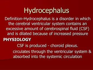
CEREBROSPINAL FLUID (CSF) • A specialized extracellular fluid produced by choroid plexi which bathes, supports and protects the brain and spinal cord.
CEREBROSPINAL FLUID (CSF) • CSF is produced by choroid plexi of lateral, 3rd and 4th ventricles. • Normal total volume of CSF is about 130-150 ml. • 30-50 ml in the ventricles, 100 ml in the subarachnoid space. • CSF is produced/resorbed by the choroid plexus/arachnoid granulations at a rate of about20 ml / hr or 500 ml/ day.
CSF Lateral ventricle Foramen of Monro 3rd ventricle Aqueduct of Sylvii 4th ventricle Foramen of Luschka and Magendi Subarachnoid space
CSF CSF then flows up to the arachnoid granulations (groups of arachnoid villi) in the superior sagittal sinus where it passes through the one-way valves into the venous blood.
HYDROCEPHALUS Increase in ventricular size due to an imbalance between the production of CSF and its drainage by the arachnoid villi.
Headache, nausea/vomiting, Cognitive impairment, decreased levelof consciousness, Papilledema, decreased vision and abducens palsies, Gait abnormalities, CLINICAL MANIFESTATIONS
CLINICAL MANIFESTATIONS • Dilated scalp veins, • Anterior fontanel wide open and bulging, increased head circumference, • Setting sun sign, • Brisk tendon reflexes, spasticity • Clonus, Babinsky sign (+)
IMAGING STUDIES • X-RAY PLAIN FILMS: • SEPARATION OF SUTURES • EROSION OF POSTERIOR CLINOIDS • INCREASED CONVOLUTIONAL MARKINGS (BEATEN CUPPER APPEAREANCE) • ULTRASOUND • CT • MRI
HYDROCEPHALUS Ventricular dilation and increased intracranial pressure due to discrepancy between CSF production and absorption DDx: Atrophy (Ex vacuo dilatation of ventricles) DDx: Hydranencephaly
HYDROCEPHALUS Commensurate dilatation of the temporal horn with the bodies of the lateral ventricles
ATROPHY Temporal horn is not dilated
HYDRANENCEPHALY • Cerebral hemispheres are virtually absent • Replaced by membranous sacs of CSF • Vascular injury: bilateral in utero internal carotid artery infarction • Large cystic mass, no cerebral cortex • Midbrain, basal ganglia, and posterior fossa are usually normal. • Shunting not indicated (unlike extreme hydrocephalus)
HYDRANENCEPHALY Shunt ???
HYDROCEPHALUS Dilatation of anterior and posterior recesses of the 3rd ventricle; decreased mamillo-pontine distance
HYDROCEPHALUS Upward bowing of the corpus callosum
HYDROCEPHALUS • Transependymal CSF resorption/migration in the periventricular white matter
HYDROCEPHALUS The situation might be compensated or noncompensated depending on the presence of transependymal CSF migration.
CLASSIFICATION Three major mechanisms account for the development of hydrocephalus: *Overproduction of CSF (communicating type), *Obstruction to CSF flow (intraventricular, non-communicating), *Decreased absorption of CSF by arachnoid villi (extraventricular, communicating)
OVERPRODUCTION OF CSF Choroid plexus papillomas: Large aggregations of choroidal fronds, Infancy, Large amounts of CSF, Atria of the lateral ventricles are the most common locations. Courtesy of Prof. A. Cila
NON-COMMUNICATING HYDROCEPHALUS HYDROCEPHALUS SECONDARY TO INTRAVENTRICULAR OBSTRUCTION OF CSF Mechanism: Obstruction to CSF flow within the ventricular system, commonat narrow points such as foramen oraqueduct *Tumors (benign and malignant), * Foraminal gliosis/stenosis, *Aqueductal stenosis/web/gliosis, *Congenital anomalies (Chiari malformations, Dandy-Walker malformations,…)
NON-COMMUNICATING HYDROCEPHALUS Lateral ventricle Foramen Monro 3rd ventricle *Stenosis/gliosis,tumor,colloid cyst Courtesy of Prof. A. Cila
NON-COMMUNICATING HYDROCEPHALUS Lateral ventricle Foramen Monro 3rd ventricle *Stenosis/gliosis,tumor,colloid cyst
NON-COMMUNICATING HYDROCEPHALUS Lateral ventricle Foramen Monro 3rd ventricle *Stenosis/gliosis,tumor,colloid cyst
NON-COMMUNICATING HYDROCEPHALUS 3rd ventricle Aqueduct of Sylvius 4th ventricle *Stenosis/gliosis,tumor,infection
NON-COMMUNICATING HYDROCEPHALUS 3rd ventricle Aqueduct of Sylvius 4th ventricle *Stenosis/gliosis,tumor,infection
NON-COMMUNICATING HYDROCEPHALUS 3rd ventricle Aqueduct of Sylvius 4th ventricle *Stenosis/gliosis,tumor,infection Courtesy of Prof. A. Cila
NON-COMMUNICATING HYDROCEPHALUS 3rd ventricle Aqueduct of Sylvius 4th ventricle *Stenosis/gliosis,tumor,infection
NON-COMMUNICATING HYDROCEPHALUS 3rd ventricle Aqueduct of Sylvius 4th ventricle *Stenosis/gliosis,tumor,infection
NON-COMMUNICATING HYDROCEPHALUS CONGENITAL MALFORMATIONS Vein of Galen Malformation 3 month-old baby boy with enlarging head circumference, and an audible cranial bruit.
NON-COMMUNICATING HYDROCEPHALUS CONGENITAL MALFORMATIONS Chiari I
NON-COMMUNICATING HYDROCEPHALUS CONGENITAL MALFORMATIONS Chiari II
NON-COMMUNICATING HYDROCEPHALUS CONGENITAL MALFORMATIONS Chiari III
NON-COMMUNICATING HYDROCEPHALUS CONGENITAL MALFORMATIONS DANDY-WALKER
COMMUNICATING HYDROCEPHALUS HYDROCEPHALUS SECONDARY TO EXTRAVENTRICULAR OBSTRUCTION OF CSF Mechanism: Decreased absorption of CSF by arachnoid villi *subarachnoid and intraventricular bleeding, *inflammatory conditions (meningitis), * tumoral seeding to subarachnoid spaces, *venous hypertension.
COMMUNICATING HYDROCEPHALUS *Subarachnoid and intraventricular bleeding
COMMUNICATING HYDROCEPHALUS *Inflammatory conditions (meningitis)
COMMUNICATING HYDROCEPHALUS *Inflammatory conditions (meningitis)
COMMUNICATING HYDROCEPHALUS * Tumoral seeding to subarachnoid spaces Courtesy of Prof. A. Cila
TREATMENT OF HYDROCEPHALUS • Persistent compression of the brain • Gliosis, cortical distortion, neuronal damage, axonal tearing along longitudinal fibers (internal capsule), and myelin destruction • Treatment is mandatory
TREATMENT OF HYDROCEPHALUS • Ventricular shunting (VP-VA shunt): CSF diversion • Endoscopic third ventriculostomy (E3V); endoscopic aqueductoplasty (EA)
TREATMENT OF HYDROCEPHALUS • Ventricular shunting (VP-VA shunt) Assess the fxn of shunt by determining the position of tip of ventriculostomy tube (around f. Monro), and the reduction in ventricular size.
TREATMENT OF HYDROCEPHALUS • Ventricular shunting complications • -Shunt malfxn-nonfxn (due to occlusion of ventricular catheter, disconnection of the shunt components, …) • -Infection (ventriculitis) • -Subdural hematomas • -Post-shunt dural fibrosis • -Slit ventricle syndrome • -Abdominal complications (ascites, pseudocyst formation, perforation of a viscus,…)