Download
1 / 29
410 likes | 784 Views
Normal Pressure Hydrocephalus ( NPH ). Date: 2005/09/27 Speaker: Int. 吳忠泰 Supervisor: V.S. 俞芹英. Outlines. Definition Epidemiology and etiology Physiology and pathophysiology Diagnosis and differential diagnosis Treatment and complication Prognosis. Definition.

E N D
Normal Pressure Hydrocephalus (NPH) Date: 2005/09/27 Speaker: Int. 吳忠泰 Supervisor: V.S. 俞芹英
Outlines • Definition • Epidemiology and etiology • Physiology and pathophysiology • Diagnosis and differential diagnosis • Treatment and complication • Prognosis
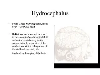
Definition • First described in 1965 by Hakim and Adams • Normal CSF pressure • Ventriculomegaly • Clinical triad: • Slowly progressive gait disorder • Impairment of mental function • Sphincteric incontinence
Epidemiology • 1 per 25’000 • Accounts for approximately 0.5-5% (up to 6%) of dementias • One of the few treatable causes of dementia • Most common in patient > 60 y/o • M > F
Etiology • Idiopathic: Elderly, unknown cause, 50% of NPH • With a preceding cause: Young • Subarachnoid hemorrhage (SAH) • Trauma • Meningitis (TB, syphilitic, etc.) • Surgery, irradiation • Storage disease (mucopolysaccharidosis)
Pathophysiology of Hydrocephalus Communicating hydrocephalus Obstructive hydrocephalus
Pathophysiology of NPH Incontinence Enlarged lateral ventricles Gait disturbance Dementia
Pathophysiology of NPH • On the basis of both dynamic and ischemic factors • Ventricular enlargement • Vascular stretching → Ischemia • Decreased compliance of ventricular wall • High pulse pressure • Barotrauma or shearing stress
Dynamics of NPH • Transmantle pressure gradient • Difference in pressure between ventricle and subarachnoid space • Gradient ↑ temporarily → Ventricle↑ • B wave (plateau) • Transient elevations of mean and pulse pressure • Water-hammer effect→ Ventricle↑ • More than 50% of time
Dynamics of NPH • Aqueductal CSF flow void • Increased CSF flow velocity • Favorable response to CSF diversion • Aqueductal CSF stroke volume • CSF pulsating back and forth through the aqueduct during systole and diastole • Favorable response to shunting • Hyperdynamic CSF flow
Dynamics of NPH • Saline infusion test • CSF resorption in NPH is abnormal • Arachnoid granulation? Arachnoidal villi? • Venous compromise • Increased transvenular resistance in superior saggital sinus cause NPH • What cause venous compromise? Microangiopathy? Deep white matter ischemia?
Ischemia of NPH • Acetazolamide challenge test • Cerebral blood flow (CBF)↑ in normal person • Failed to cause CBF↑ in NPH p’t • Indicate the arterioles are already maximally dilated because of ischemia • CSF diversion → CBF improve and response to acetazolamide
Ischemia of NPH • Compensatory CSF flow • Periventricular white matter • Increased interstitial fluid • Loss of parenchymal compliance
Pathophysiology of NPH • Dynamic • Hyperdynamic CSF flow • Impaired CSF resorption • Ischemic • Reduced CBF • Periventricular white matter lesion
Diagnosis • Clinical symptoms and signs • Gait disturbance • Dementia • Urinary incontinence • The moment when highly suspect NPH !! • Image • MRI (T2WI) with CSF flow study • CT with lumbar puncture
Image Findings - CT • Ventriculomegaly • Sulcal atrophy • Ventriculosulcal disproportion • Can DDx with other dementia syndromes
Image Findings - CT • Rounded frontal and temporal horns • Periventricular lucency • Transependymal CSF flow • Corpus callosum thinning
Image Findings - MR • The same as CT • Temporal horn out of proprotion to hippocampal atrophy • Corpus callosum bowed upward
Image Findings - MR • Periventricular lesions in T2WI • Transependymal CSF flow • Deep white matter damage
Image Findings - MR • Aqueductal flow void sign • Jet sign • A jet of turbulent CSF flow on the distal aqueduct • Predictive of shunt responsiveness
Image Studies - MR • CSF flow study • Aqueductal stroke volume • Increased velocity (hyperdynamic flow) • VV/ICV ratio • VV/ICV ratio > 30% (in 13 of 14 pts) (VV: ventricular volume; ICV: intracranial CSF space volume) • MRS • Intraventricular lactate peaks (ischemia)
Differential Diagnosis • Dementia syndromes • Alzheimer’s disease • Hydrocephalus ex vacuo • Intraventricular lactate • Parkinsonism • Parkinson’s disease • Periventricular leukomalacia
Treatment • Surgical shunting • VP shunt • Lumbar puncture • Miller Fisher test: Gait assessment before and after 30mL CSF drainage (high rate of false negative) • Continuous CSF drainage of 200 mL per day for 3-5 days
Complications of shunt • Infection: S. aureus, S. epidermidis • Subdural hematoma • Shunt obstruction • Low pressure state • Epilepsy • Pneumocephalus • Ascites
Prognosis • Response rate for shunt • 50-70% with known preceding cause • 30% with idiopathic group • Non-selective patient • 1/3 improve, 1/3 arrest, 1/3 deteriorate
Prognosis • Positive response to shunting: • Absence of central atrophy or ischemia • Gait apraxia as dominant symptoms • Prominent CSF flow void (stroke volume > 42 mL) • Known history of cause (nonidiopathic type)
References • Raymond D. Adams, Maurice Victor. Principles of Neurology, 5th edition: 545-6 • Roger N. Rosenberg et al. The Clinical Neuroscience – Neurology/ Neurosurgery: 1205-19 • Kenneth W. Lindsay, Ian Bone. Neurology and Neurosurgery Illustrated, 4th edition: 128-9, 370-3 • Anne G. Osborn et al. Pocket Radiologist – Brain Top 100 Diagnosis: 228-30 • William G. Bradley. Normal Pressure Hydrocephalus: New Concepts on Etiology and Diagnosis. AJNR 2000; 21: 1586-90 • eMedicine: http://www.emedicine.com/neuro/topic277.htm http://www.emedicine.com/radio/topic479.htm