Download
1 / 1
20 likes | 155 Views
Spontaneous plasticity in brain sensory maps in rats after focal stroke revealed by repeated fMRI. (and how to get better results by using fewer animals in longitudinal brain imaging studies). Denise Duricki, Toby Wood, Camilla Simmons, Michel Bernanos,

E N D
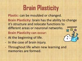
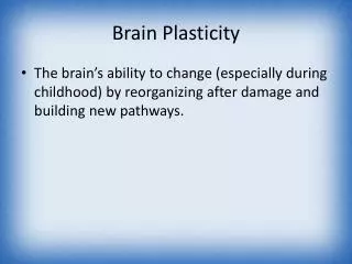
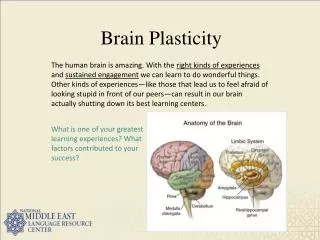
Spontaneous plasticity in brain sensory maps in rats after focal stroke revealed by repeated fMRI. (and how to get better results by using fewer animals in longitudinal brain imaging studies) Denise Duricki, Toby Wood, Camilla Simmons, Michel Bernanos, Steve Williams, Diana Cash & Lawrence Moon Wolfson Centre for Age-Related Diseases and Department of Neuroimaging, King’s College London Results (p<0.001) Abstract Results (p<0.01) As part of a study evaluating a novel stroke therapy, we have optimised a method for repeatedly imaging brain activity in rats. Sensory cortex activity was visualised indirectly using BOLD-fMRI (Blood Oxygen Level-Dependent Functional Magnetic Resonance Imaging) during paw stimulation. Previously, adequate signal was only obtained under terminal anaesthesia, but new protocols allow multiple sessions of imaging under transient anaesthesia using medium-dose alpha-chloralose (de Alonso et al 2011). We now show that the same rat can be imaged prior to stroke and 1 and 8 weeks after stroke. This allows powerful longitudinal statistics to be applied. We report changes in sensory cortex activation over time after stroke in response to paw stimulation. Pre-stroke: strong sensory cortex activation during paw stimulation under optimised alpha-chloralose anaesthesia Statistical comparison; pre-stroke vs. one week Loss of signal in affected hemisphere; opposite hemisphere more responsive to stimulation of less-affected paw Stimulation of dominant paw Stimulation of less-dominant paw Stimulation of dominant paw Stimulation of less-dominant paw Introduction to BOLD fMRI • Increased regional brain activity involves increased oxygen consumption; thus more conversion of oxyhaemoglobin to deoxyhaemoglobin. • OxyH and DeoxyH have different magnetic properties. • A magnetic field is used to align the protons in molecules in the brain ( ) but local magnetic fields (e.g., due to oxyH or deoxyH) affect alignment ( ). • Non aligned protons are detected by pulsing the tissue with a radiowave, causing them to emit energy that allows their location to be determined. • Stimulation of less-dominant paw causes widespread activation and suppression. • Need to keep rats warm, monitor respiration, give fluids. • New anaesthetic regime works well. • Stimulation of dominant paw causes focal activation of sensory cortex. Statistical comparison: week 1 vs. week 8 Recovery of signal in affected hemisphere; normalisation in opposite hemisphere One week post-stroke: Loss of sensory cortex activation in affected hemisphere Stimulation of dominant paw Stimulation of dominant paw Stimulation of less-dominant paw Stimulation of less-dominant paw Choice of anaesthetic is crucial α-chloralose Domitor Our Domitor result BOLD-fMRI with paw stimulation causes activation of sensory cortex. (Left) Alpha-chloralose provides the best signal but at this dose is a terminal anaesthetic; (Middle) Domitor (medetomidine) gives less signal but allows recovery (Weber et al 2006). (Right) Our preliminary findings found only poor signal with Domitor. • As expected, total loss of signal in infarct area when affected paw is stimulated. • Heightened, widespread response in opposite hemisphere to stimulation of less-dominant paw • Affected hemisphere also shows some loss of signal in dorsal striatum and in opposite hemisphere Eight weeks post-stroke: Recovery of sensory cortex activation in affected hemisphere; normalisation of opposite hemisphere Conclusions 1. The novel anaesthetic regime described by de Alonso et al (2011) is very suitable for longitudinal (repeated measures) fMRI in rats. 2. A strong signal is obtained in sensory cortex during stimulation of either paw. 3. The dominant paw has an intense response focused on sensory cortex. 4. Stroke causes a transient decrease in signal in the affected side at 1 week and a spontaneous increase in peri-lesional activity occurs by 8 weeks. Stimulation of dominant paw Stimulation of less-dominant paw BOLD-fMRI with terminal alpha-chloralose requires many animals per timepoint (n = 60 for 5 time points) in order to detect changes in sensory cortex activation after spinal cord injury (Endo et al., 2007) which wastes animals and involves greater interanimal variability. But just at the right moment, during our pilot studies, de Celis Alonso et al (2011) published a report showing medium dose alpha-chloralose allows recovery after BOLD-fMRI in rats with excellent signal during paw stimulation. We now report positive findings using this method. Methods • Recovery of signal in peri-infarct area following stimulation of affected paw. • Dorsal striatum less active following stimulation of affected paw. • Following stimulation of the less-affected paw, activation is less widespread. • Adult rats were imaged either naive or one or eight weeks after focal stroke. • Stroke was induced by microinjection of • the vasoactive peptide endothelin-1 into • the sensorimotor cortex of adult rats: this • restricts blood flow, causing an ischemic • lesion and a persistent deficit in forelimb • function (Soleman et al., 2010). • Rats were anaesthetised using alpha chloralose as described in de Celis Alonso et al. (2011) and BOLD-fMRI images were acquired during stimulation of the left or right paw in a pseudorandom order. • 7 Tesla VMRIS MRI suite at James Black Institute, IOP. • Images consist of “voxels” where each point represents • the probability that this point was active on an “on” pulse. References: de Celis Alonso et al (2011) On the use of alpha-chloralose for repeated BOLD fMRI measurements in rats. Journal of Neuroscience Methods. 195: 236-240. Funding: Thanks to the RCUK, MRC, British Pharmacological Society (BPS)’s Integrative Pharmacology Fund, funding from the Centre for Integrative Biomedicine (a pre-CASE award and a Systems Biology grant) and a Capacity Building Award in Integrative Mammalian Biology funded by the BBSRC, BPS, HEFCE, Knowledge Transfer Partnerships, MRC and Scottish Funding Council. 3Hz, 2mA, 0.3s off/on (pseudo random) stimulation of the left forepaw