Download
1 / 86
940 likes | 1.47k Views
6 Osseous Tissue and Bone Structure. Section 1: An Introduction to Bones. Learning Outcomes 6.1 Classify bones according to their shapes, identify the major types of bone markings, and explain the functional significance of surface features.

E N D
6 Osseous Tissue and Bone Structure
Section 1: An Introduction to Bones Learning Outcomes 6.1 Classify bones according to their shapes, identify the major types of bone markings, and explain the functional significance of surface features. 6.2 Identify the parts of a typical long bone, and describe its internal structures. 6.3 Identify the cell types in bone, and list their major functions. 6.4 Compare the structures and functions of compact bone and spongy bone.
Learning Outcomes 6.5 Explain the process of appositional bone growth. 6.6 Explain the mechanisms of endochondral ossification. 6.7 Explain the mechanisms of intramembranous ossification. 6.8CLINICAL MODULE Discuss various abnormalities of bone formation and growth. Section 1: An Introduction to Bones
Skeletal system components Bones (206 total) Divisions Axial skeleton (126 bones) Bones of skull, thorax, and vertebral column Form longitudinal axis of body Appendicular skeleton (80 bones) Bones of the limbs and girdles that attach them to the axial skeleton Cartilages Ligaments and other connective tissues Section 1: An Introduction to Bones
Figure 6 Section 1 1 Axial Skeleton (126 Bones) The axial skeleton consists of the bones of the skull, thorax, and vertebral column. These elements form the longitudinal axis of the body. Appendicular Skeleton (80 Bones) The adult skeletal system, which can be divided into the axial skeleton and the appendicular skeleton The appendicular skeleton includes the bones of the limbs and the pectoral and pelvic girdles that attach the limbs to the axial skeleton.
Functions of the skeletal system Support (support for body, attachment for soft tissues) Storage of minerals (calcium and phosphate) Calcium most abundant mineral in body (~2–4 lb) 98% stored in bones Blood cell production (all formed elements of blood) Protection (delicate tissues and organs surrounded by bone) Leverage (act as levers with skeletal muscles to move body) Section 1: An Introduction to Bones
Module 6.1: Bone classification Six categories based on shape Flat bones Thin, roughly parallel surfaces Examples: cranial bones, sternum Sutural bones (Wormian bones) Irregular bones formed between cranial bones Number, size, and shape vary Long bones Relatively long and slender Examples: various bones of the limbs
Six categories based on shape (continued) Irregular bones Complex shapes Examples: vertebrae, bones of pelvis, facial bones Sesamoid bones Small, flat, and somewhat shaped like sesame seed Develop in tendons of knee, hands, and feet Individual variation in location and number Short bones Small and boxy Examples: bones of the wrist (carpals) and ankles (tarsals) Module 6.1: Bone classification
Bone surface features Also known as bone markings External and internal features related to functions Elevations/projections for tendon and ligament attachment Depressions/grooves/tunnels for blood vessels or nerves to lie alongside or penetrate Module 6.1: Bone classification
Skull surface features Canal or meatus (large passageway) Process (projection or bump) Sinus (chamber within bone, usually filled with air) Foramen (small rounded passageway) Fissure (elongated cleft or gap) Module 6.1: Bone classification
Humerus surface features Head (expanded proximal end that forms part of joint) Tubercle (small, rounded projection) Sulcus (deep, narrow groove) Tuberosity (small, rough projection; may occupy broad area) Diaphysis (shaft; elongated body) Trochlea (smooth, grooved articular process) Condyle (smooth, rounded articular process) Module 6.1: Bone classification
Femur surface features Trochanter (large, rough projection) Head Neck (narrow connection between head and diaphysis) Diaphysis Facet (small, flat articular surface) Condyle Module 6.1: Bone classification
Pelvis surface features Crest (prominent ridge) Fossa (shallow depression or recess) Line (low ridge; more delicate than crest) Spine (pointed or narrow process) Ramus (extension that makes angle with rest of structure) Module 6.1: Bone classification
Module 6.1 Review a. Define surface feature. b.Identify the six broad categories for classifying a bone according to shape. c.Compare a tubercle with a tuberosity.
Module 6.2: Typical long bone structure Long bone features Epiphysis (expanded ends) Consist largely of spongy bone (trabecular bone) Network of struts and plates Resists forces from various directions and directs body weight to diaphysis and joints Outer covering of compact bone Strong, organized bone Articular cartilage Covers portions of epiphysis that form articulations Avascular and receives resources from synovial fluid
Long bone features (continued) Metaphysis (connects epiphysis to shaft) Diaphysis (shaft) Contains medullary cavity (marrow cavity) Filled with marrow Red bone marrow (red blood cell production) Yellow bone marrow (adipose storage) Module 6.2: Typical long bone structure
Figure 6.2 1 - 2 Coronal sections through a right femur, showing the boundaries of a long bone’s major regions, plus the bone’s internal organization and how it distributes the forces applied to the bone Body weight (applied force) The epiphysis consists largely of spongy bone, also called trabecular bone. Spongy bone consists of an open network of struts and plates that resembles latticework with a thin covering, or cortex, of compact bone. The epiphysis (e-PIF-i-sis) is an expanded area found at each end of the bone. The metaphysis (me-TAF-i-sis; meta, between) is a narrow zone that connects the epiphysis to the shaft of the bone. The wall of the diaphysis consists of a layer of compact bone. Tension on lateral side of shaft The medullary cavity (medulla, innermost part), or marrow cavity, is a space within the hollow shaft. In life, it is filled with bone marrow, a highly vascular tissue. Red bone marrow is highly vascular and involved in the production of blood cells. Yellow bone marrow is adipose tissue important in the storage of energy reserves. The diaphysis (shaft) is long and tubular. Compression on medial side of shaft Metaphysis Epiphysis
Bone vasculature Growth and maintenance requires extensive blood supply Vascular features Nutrient artery/vein (commonly one each/bone) Nutrient foramen (tunnel providing access to marrow cavity) Also supplies osteons of compact bone with blood Metaphyseal artery/vein Carry blood to/from metaphysis Connects to epiphyseal arteries/veins Module 6.2: Typical long bone structure
Figure 6.2 3 A longitudinal section of the humerus, showing the extensive|network of blood vessels in long bones An articular cartilage covers portions ofthe epiphysis that articulate with other bones. The cartilage is avascular, and itrelies primarily on diffusion from thesynovial fluid to obtain oxygen andnutrients and eliminate wastes. Epiphyseal arteryand vein The metaphyseal artery (red) andmetaphyseal vein (blue) carry blood toand from the area of the metaphysis and tothe epiphysis through epiphyseal arteriesand veins. Metaphysis Most bones have only onenutrient artery (shown inred) and one nutrient vein(shown in blue), but a fewbones, including the femur,have more than one of each. Periosteum Compactbone Medullarycavity A nutrient foramen is a tunnel that penetrates thediaphysis and providesaccess for the nutrient arteryand/or vein. Branches ofthese large vessels supplythe osteons of thesurrounding compact bonebefore entering andsupplying the tissues of themedullary cavity. Metaphysealartery and vein Metaphysis
Periosteum features Smaller blood vessels (supply superficial osteons) Lymphatic vessels (collect lymph from bone and osteons) Sensory nerves (innervate diaphysis, medullary cavity, and epiphyses) Module 6.2: Typical long bone structure
Module 6.2 Review a.List the major parts of a long bone. b. Describe the function of the medullary cavity. c.If articular cartilage is avascular, how is it nourished?
Module 6.3: Bone tissue Four bone cell types Osteocytes (osteo-, bone + cyte, cell) Mature bone cells that cannot divide Most numerous bone cell type Maintain protein and mineral content of adjacent matrix Dissolve matrix to release minerals Rebuild matrix to deposit mineral crystals Occupy lacunae (pocket) Separated by layers of matrix (lamellae) Connected with canaliculi
Four bone cell types (continued) Osteoblasts (blast, precursor) Produce new bony matrix (osteogenesis or ossification) Begins with release of proteins and other organic components to produce unmineralized matrix (= osteoid) Then assists in depositing calcium salts to convert osteoid to bone Become osteocytes once surrounded by bony matrix Module 6.3: Bone tissue
Figure 6.3 1 - 2 The structures of osteocytes and osteoblasts within a long bone The layers of matrix are called lamellae (lah-MEL-lē; singular, lamella, a thin plate). Narrow passageways called canaliculi penetrate the lamellae, radiating through the matrix and connecting lacunae to one another and to various blood vessels that supply nutrients. Osteocytes account for most of the cell population in bone. Each osteocyte occupies a lacuna, a pocket sandwiched between layers of matrix. Osteocytes cannot divide, and a lacuna never containsmore than one osteocyte. Osteoblast Osteoid
Four bone cell types (continued) Osteoprogenitor cells (progenitor, ancestor) Mesenchymal (stem) cells that produce cells that differentiate into osteoblasts Important in fracture repair Locations Inner lining of periosteum Lining endosteum in medullary cavity Lining passageways containing blood vessels Module 6.3: Bone tissue
Four bone cell types (continued) Osteoclasts (clast, to break) Remove and remodel bone matrix Giant cells with 50+ nuclei Derived from same stem cells as macrophages Release acids and proteolytic enzymes to dissolve matrix and release stored minerals = Osteolysis (lysis, loosening) Module 6.3: Bone tissue
Figure 6.3 3 - 4 The structures of osteocytes and osteoblasts within a long bone Endosteum Osteoprogenitor cell Osteoclast
Module 6.3: Bone tissue Bone matrix Collagen fibers account for ~1/3 bone weight Provide flexibility Calcium phosphate (Ca3(PO4)2) accounts for ~2/3 bone weight Interacts with calcium hydroxide (Ca(OH)2) to form crystals of hydroxyapatite (Ca10(PO4)6(OH)2) salts Incorporates other salts (calcium carbonate, CaCO3) and ions (Na, Mg2, F) Provides strength
Module 6.3 Review a.Define osteocyte, osteoblast, osteoprogenitor cell, and osteoclast. b.How would the compressive strength of a bone be affected if the ratio of collagen to hydroxyapatite increased? c.If osteoclast activity exceeds osteoblast activity in a bone, what would be the effect on the bone?
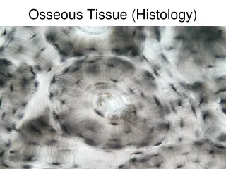
Module 6.4: Compact and spongy bone Compact bone Functional unit is osteon Organized concentric lamellae around a central canal Osteocytes (in lacunae) lie between lamellae Central canal contains small blood vessels Canaliculi connect lacunae with each other and central canal Strong along its length
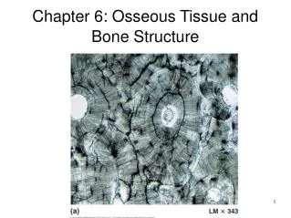
Figure 6.4 1 - 2 The structure of compact bone, as shown in the shaft of a long bone Capillary and venule Central canal Concentric lamellae Canaliculi radiating through the lamellae interconnect the lacunae of the osteons with one another and with the central canal. Endosteum Central canal Periosteum Circumferential lamellae Osteon Vein Interstitial lamellae Artery Compact bone LM x 375 The osteocytes occupy lacunae that lie between the lamellae. In preparing this micrograph, a small piece of bone was ground down until it was thin enough to transmit light. In this process, the lacunae and canaliculi are filled with bone dust, and thus appear black. Central canal Perforating canal
Module 6.4: Compact and spongy bone Typical long bone organization Periosteum (outermost layer) Compact bone (outer bone tissue layer) Circumferential lamellae (circum-, around + ferre, to bear) Outer and inner surfaces of compact bone layer Interstitial lamellae Fill spaces between osteons Osteons Contain central canals (parallel to bone surface) Connected by perforating canals (perpendicular) Spongy bone (innermost layer)
Spongy bone Located where bones not heavily stressed or in many directions Lamellae form struts and plates (trabeculae) creating an open network Reduces weight of skeleton No blood vessels in matrix Nutrients reach osteons through canaliculi open to trabeculae surfaces Module 6.4: Compact and spongy bone
Figure 6.4 3 – 4 The structure of spongy bone, as shown in the head of the femur Trabeculae of spongy bone Canaliculi opening on surface Endosteum Lamellae
Module 6.4 Review a. Define osteon. b.Compare the structures and functions of compact bone and spongy bone. c.A sample of bone has lamellae that are not arranged in osteons. Is the sample more likely from the epiphysis or from the diaphysis?
Module 6.5: Appositional bone growth Appositional bone growth Increases bone diameter of existing bones Does not form original bones Osteoprogenitor cells differentiate into osteoblasts that add bone matrix under periosteum Adds successive layers of circumferential lamellae Trapped osteoblasts become osteocytes Deeper lamellae recycled and replaced by osteons Osteoclasts remove matrix at inner surface to enlarge medullary cavity
Figure 6.5 1 Increase in bone diameter resulting from appositional growth Additional circumferential lamellae are deposited, and the bone continues to increase in diameter. Periosteum
Figure 6.5 2 Enlargement of the medullary cavity with increased bone diameter resulting from appositional growth Bone matrix is removed by osteoclasts Bone deposited by superficial osteoblasts Infant Child Adult Young adult
Periosteum Two layers Fibrous outer layer Cellular inner layer Functions Isolate bone from surrounding tissues Route for blood and nervous supply Actively participate in bone growth and repair Module 6.5: Appositional bone growth
Perforating fibers Created by osteoblasts in periosteum cellular layer Strongly connect tendons, ligaments, and joint capsules to bone through periosteum Module 6.5: Appositional bone growth
Figure 6.5 3 Structure of the periosteum Circumferential lamellae Fibrous layer of periosteum Cellular layer of periosteum Canaliculi Osteocyte in lacuna Perforating fibers
Endosteum Incomplete cellular layer lining medullary cavity Covers spongy bone and lines central canals Consists of simple layer of osteoprogenitor cells Where incomplete, osteoclasts and osteoblasts remodel matrix Module 6.5: Appositional bone growth
Figure 6.5 4 Structure of the endosteum Endosteum Osteoclast Circumferential lamella Osteocyte Osteoprogenitor cell Osteoid Osteoblast
Module 6.5 Review a.Define appositional growth. b.Distinguish between the periosteum and the endosteum. c.As a bone increases in diameter, what happens to the medullary cavity?
Module 6.6: Endochondral ossification Initial bone formation in embryo begins with cartilage Replaced by bone through endochondral (endo-, inside + chondros, cartilage) ossification Uses cartilage as small model Bone grows in diameter and length Diameter growth involves appositional bone deposition Animation: Early Endochondral Ossification
Steps of endochondral ossification In shaft, chondrocytes enlarge and matrix ossifies Chrondrocytes die, leaving cavities within cartilage Blood vessels grow around cartilage edge and osteoblasts form to create a superficial layer of bone Blood vessels penetrate central region Allow entering fibroblasts to change into osteoblasts Spongy bone produced (primary ossification center) and spreads toward bone ends Module 6.6: Endochondral ossification
Steps of endochondral ossification (continued) Medullary cavity created as cartilage replaced by osseous tissue Bone grows in length and diameter Secondary ossification centers form as capillaries and osteoblasts migrate into epiphyses Epiphyses fill with spongy bone Only articular cartilage (on epiphyses) and epiphyseal cartilage (in metaphysis) remain Module 6.6: Endochondral ossification
Figure 6.6 1 – 6 The process of endochondral ossification Hyaline cartilage Articular cartilage Spongy bone Epiphysis Enlarging chondrocytes within calcifying matrix Metaphysis Epiphysis Epiphyseal cartilage Medullary cavity Medullary cavity Periosteum Blood vessel Primary ossification center Diaphysis Compact bone Superficial bone Diaphysis Spongy bone Formation of an epiphyseal cartilage between epiphysis and diaphysis Metaphysis Bone formation Secondary ossification center Hyaline cartilage Further growth in length and diameter Enlargement of chondrocytes Formation of superficial layer of bone Production of spongy bone at a primary ossification center Formation of secondary ossification centers
Steps of endochondral ossification (continued) Bone grows in length at epiphyseal cartilage Chondrocytes actively produce more cartilage on epiphysis side Osteoblasts actively replace cartilage with bone on shaft side As long as both processes are equally active, bone lengthening continues At puberty, hormones increase bone growth and epiphyseal cartilage is replaced Leaves epiphyseal line in adults Module 6.6: Endochondral ossification