Download
1 / 81
830 likes | 850 Views
6 Osseous Tissue and Bone Structure. An Introduction to the Skeletal System. Learning Outcomes 6-1 Describe the primary functions of the skeletal system.

E N D
6 Osseous Tissue and Bone Structure
An Introduction to the Skeletal System • Learning Outcomes • 6-1 Describe the primary functions of the skeletal system. • 6-2 Classify bones according to shape and internal organization, giving examples of each type, and explain the functional significance of each of the major types of bone markings. • 6-3 Identify the cell types in bone, and list their major functions.
An Introduction to the Skeletal System • Learning Outcomes • 6-4 Compare the structures and functions of compact bone and spongy bone. • 6-6 Describe the remodeling and homeostatic mechanisms of the skeletal system. • 6-9 Describe the types of fractures, and explain how fractures heal.
An Introduction to the Skeletal System • The Skeletal System • Includes: • Bones of the skeleton • Cartilages, ligaments, and connective tissues
6-1 Functions of the Skeletal System • Five Primary Functions of the Skeletal System • Support • Storage ___________ (calcium) and Lipids (yellow marrow) • _____________Production (red marrow) • Protection • Leverage (_______________)
6-2 Classification of Bones • Bones • Are classified by: • 1. • 2. • 3.
6-2 Classification of Bones Six Bone Shapes Sutural bones ____________ bones Short bones Flat bones Long bones ______________bones
Figure 6-1 A Classification of Bones by Shape Flat Bones Sutural Bones Sutures External table Parietal bone Sutural bone Internal table Diploë (spongy bone) Long Bones Irregular Bones Vertebra Humerus Short Bones Sesamoid Bones Patella Carpal bones
6-2 Classification of Bones • Sutural Bones • Small, irregular bones • Found __________ the ______________ the skull • Irregular Bones • Have_____________ shapes • Examples: spinal __________, pubic bones
Figure 6-1a A Classification of Bones by Shape Sutural Bones Sutures Sutural bone
Figure 6-1b A Classification of Bones by Shape Irregular Bones Vertebra
6-2 Classification of Bones • Short Bones • _______ and thick, “potato”-shaped • Examples: ankle and ___________ bones • Flat Bones • __________ with parallel surfaces • Found in the skull, sternum, ribs, and scapulae
Figure 6-1c A Classification of Bones by Shape Short Bones Carpal bones
Figure 6-1d A Classification of Bones by Shape Flat Bones External table Parietal bone Internal table Diploë (spongy bone)
6-2 Classification of Bones Long Bones Long and thin Found in arms, legs, hands,_______,________, and toes Sesamoid Bones Small and flat Develop inside _____________ near joints of knees, hands, and feet. Example: ________________
Figure 6-1e A Classification of Bones by Shape Long Bones Humerus
Figure 6-1f A Classification of Bones by Shape Sesamoid Bones Patella
6-2 Classification of Bones • Bone Markings • Depressions or grooves • Along bone surface • Elevations or projections • Where tendons and ligaments attach • At articulations with other bones • Tunnels • Where blood and nerves enter bone
Table 6-1 An Introduction to Bone Markings Trochanter Sinus Head Neck Head Tubercle Crest Sulcus Neck Fossa Foramen Fissure Spine Process Tuberosity Ramus Line Facet Foramen Fossa Ramus Tubercle Trochlea Skull Pelvis Condyle Condyle Femur Humerus Let’s look at some examples!
6-2 Classification of Bones • Structure of a Long Bone • Diaphysis • The shaft • A heavy wall of __________ bone, or dense bone • A central space called _______________ (marrow) cavity • Epiphysis • Wide part at each end • Articulation with other bones • Mostly spongy (_____________) bone • Covered with compact bone (cortex) • Metaphysis • Where diaphysis and epiphysis meet
Figure 6-2a Bone Structure Epiphysis Spongy bone Metaphysis Anatomy of a bone -Coloring Compact bone Diaphysis (shaft) Medullary cavity Metaphysis Epiphysis The structure of a representative long bone (the femur) in longitudinal section
6-2 Classification of Bones • Structure of a Flat Bone • The parietal bone of the skull • Resembles a sandwich of spongy bone • Between two layers of compact bone • Within the cranium, the layer of spongy bone between the compact bone is called the diploë
Figure 6-2b Bone Structure Cortex (compact bone) Diploë (spongy bone) The structure of a flat bone (the parietal bone)
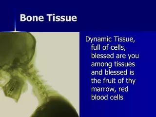
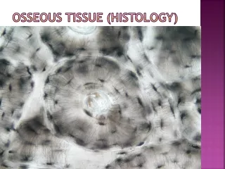
6-3 Bone (Osseous) Tissue • Bone (_____________) Tissue • Dense, supportive connective tissue • Contains specialized cells • Produces solid matrix of calcium salt deposits • Around collagen fibers
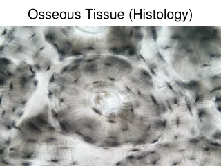
6-3 Bone (Osseous) Tissue • Characteristics of Bone Tissue • Dense matrix, containing: • Deposits of calcium salts • Osteocytes (bone cells) within lacunae organized around blood vessels • Canaliculi • Form pathways for blood vessels • Exchange nutrients and wastes
6-3 Bone (Osseous) Tissue • Characteristics of Bone Tissue • Periosteum • Covers outer surfaces of bones • Consists of outer fibrous and inner cellular layers
6-3 Bone (Osseous) Tissue • Bone Matrix • Minerals • Two thirds of bone matrix is calcium___________, Ca3(PO4)2 • Reacts with calcium hydroxide, Ca(OH)2 • To form crystals of _________________, Ca10(PO4)6(OH)2 • Which incorporates other calcium salts and ions
6-3 Bone (Osseous) Tissue • Bone Matrix • Matrix Proteins • One third of bone matrix is protein fibers (__________)
6-3 Bone (Osseous) Tissue • Bone Cells • Make up only 2% of bone mass • Bone contains four types of cells • 1. • 2. • 3. • 4.
Figure 6-3 Types of Bone Cells Canaliculi Osteocyte Matrix Matrix Osteoid Osteoblast Osteoblast: Immature bone cell that secretes organic components of matrix Osteocyte: Mature bone cell that maintains the bone matrix Osteoprogenitor cell Osteoclast Matrix Medullary cavity Medullary cavity Endosteum Osteoprogenitor cell: Stem cell whose divisions produce osteoblasts Osteoclast: Multinucleate cell that secretes acids and enzymes to dissolve bone matrix
6-3 Bone (Osseous) Tissue • Osteocytes • Mature bone cells that maintain the bone matrix • Live in ___________________ • Are between layers (lamellae) of matrix • Connect by cytoplasmic extensions through ____________ in lamellae • Do not divide • Two major functions of osteocytes • To maintain ____________ and ___________content of matrix • To help repair damaged bone
Figure 6-3 Types of Bone Cells Osteocyte Matrix Canaliculi Osteocyte: Mature bone cell that maintains the bone matrix
6-3 Bone (Osseous) Tissue • Osteoblasts • Immature bone cells that secrete matrix compounds (______________________) • Osteoid — matrix produced by osteoblasts, but not yet calcified to form bone • Osteoblasts surrounded by bone become ________________
Figure 6-3 Types of Bone Cells Osteoid Matrix Osteoblast Osteoblast: Immature bone cell that secretes organic components of matrix
6-3 Bone (Osseous) Tissue • Osteoprogenitor Cells • Mesenchymal stem cells that divide to produce osteoblasts • Located in endosteum, the inner cellular layer of periosteum • Assist in fracture repair
Figure 6-3 Types of Bone Cells Osteoprogenitor cell Medullary cavity Endosteum Osteoprogenitor cell: Stem cell whose divisions produce osteoblasts
6-3 Bone (Osseous) Tissue • Osteoclasts • Secrete acids and __________________ enzymes • _______________, multinucleate cells • Dissolve bone matrix and release stored minerals (_______________________) • Derived from stem cells that produce macrophages
Figure 6-3 Types of Bone Cells Osteoclast Matrix Medullary cavity Osteoclast: Multinucleate cell that secretes acids and enzymes to dissolve bone matrix
6-3 Bone (Osseous) Tissue • Homeostasis • Bone building (by osteoblasts) and bone recycling (by osteoclasts) must balance • More breakdown than building, bones become weak • Exercise, particularly weight-bearing exercise, causes osteoblasts to build bone
6-4 Compact Bone and Spongy Bone • The Structure of Compact Bone • Osteon is the basic unit • Osteocytes are arranged in concentric lamellae • Around a central canal containing blood vessels • Perforating canals • Perpendicular to the central canal • Carry blood vessels into bone and marrow
6-4 Compact Bone and Spongy Bone • The Structure of Compact Bone • Circumferential Lamellae • Lamellae wrapped around the long bone • Bind osteons together
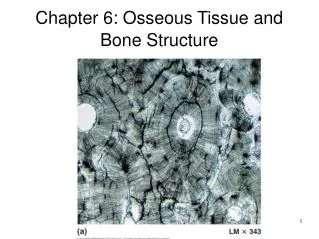
Figure 6-4a The Histology of Compact Bone Canaliculi Concentric lamellae Central canal Osteon Lacunae Osteon LM 343 A thin section through compact bone. By this procedure the intact matrix making up the lamellae appear white, and the central canal, lacunae, and canaliculi appear black due to the presence of bone dust.
Figure 6-4b The Histology of Compact Bone Osteon Lacunae Central canals Lamellae Osteons SEM 182 Several osteons in compact bone.
Figure 6-5a The Structure of Compact Bone Venule Circumferential lamellae Capillary Periosteum Osteons Perforating fibers Interstitial lamellae Concentric lamellae Trabeculae of spongy bone (see Fig.6–6) Vein Artery Perforating canal Arteriole Central canal The organization of osteons and lamellae in compact bone
Figure 6-5a The Structure of Compact Bone Central canal Concentric lamellae Endosteum The organization of osteons and lamellae in compact bone
Figure 6-5b The Structure of Compact Bone Collagen fiber orientation The orientation of collagen fibers in adjacent lamellae
6-4 Compact Bone and Spongy Bone • The Structure of Spongy Bone • Does not have ___________________ • The matrix forms an open network of trabeculae • Trabeculae have no blood vessels • The space between trabeculae is filled with ___________marrow • Which has blood vessels • Forms red blood cells • And supplies nutrients to osteocytes • Yellow bone marrow • In some bones, spongy bone holds yellow bone marrow • Is yellow because it stores______________
Figure 6-6 The Structure of Spongy Bone Trabeculae of spongy bone Canaliculi opening on surface Endosteum Lamellae
6-4 Compact Bone and Spongy Bone • Weight-Bearing Bones • The femur transfers weight from hip joint to knee joint • Causing tension on the lateral side of the shaft • And compression on the medial side