Download

1 / 111
1.19k likes | 1.74k Views
Osseous Tissue and Bone Structure. BIOL241 “Lecture 6” (2 nd ) 3 rd Week INTERCONNECTEDNESS. Topics:. Skeletal cartilage Structure and function of bone tissues Types of bone cells Structures of the two main bone tissues Bone membranes Bone formation

E N D
Osseous Tissue and Bone Structure BIOL241 “Lecture 6” (2nd) 3rd Week INTERCONNECTEDNESS
Topics: • Skeletal cartilage • Structure and function of bone tissues • Types of bone cells • Structures of the two main bone tissues • Bone membranes • Bone formation • Minerals, recycling, and remodeling • Hormones and nutrition • Fracture repair • The effects of aging
The Skeletal System • Skeletal system includes: • bones of the skeleton • cartilages, ligaments, and connective tissues
Skeletal Cartilage • Contains no blood vessels or nerves • Surrounded by the perichondrium (dense irregular connective tissue) that resists outward expansion • Three types – hyaline, elastic, and fibrocartilage
Hyaline Cartilage • Provides support, flexibility, and resilience • Is the most abundant skeletal cartilage • Is present in these cartilages: • Articular – covers the ends of long bones • Costal – connects the ribs to the sternum • Respiratory – makes up larynx, reinforces air passages • Nasal – supports the nose
Elastic Cartilage • Similar to hyaline cartilage, but contains elastic fibers • Found in the external ear and the epiglottis
Fibrocartilage • Highly compressed with great tensile strength • Contains collagen fibers • Found in menisci of the knee and in intervertebral discs
Growth of Cartilage • Appositional – cells in the perichondrium secrete matrix against the external face of existing cartilage • Interstitial – lacunae-bound chondrocytes inside the cartilage divide and secrete new matrix, expanding the cartilage from within • Calcification of cartilage occurs • During normal bone growth • During old age
Bones and Cartilages of Homo sapiens Figure 6.1
Functions of the Skeletal System • Support • Storage of minerals (calcium) • Storage of lipids (yellow marrow) • Blood cell production (red marrow) • Protection • Leverage (force of motion)
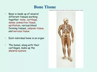
Bone (Osseous) Tissue • Supportive connective tissue • Very dense • Contains specialized cells • Produces solid matrix of calcium salt deposits and collagen fibers
Characteristics of Bone Tissue • Dense matrix, containing: • deposits of calcium salts • osteocytes within lacunae organized around blood vessels • Canaliculi: • form pathways for blood vessels • exchange nutrients and wastes
Characteristics of Bone Tissue • Periosteum: • covers outer surfaces of bones • consist of outer fibrous and inner cellular layers • Contains osteblasts responsible for bone growth in thickness • Endosteum • Covers inner surfaces of bones
Bone Matrix • Solid ground is made of mineral crystals • ⅔ of bone matrix is calcium phosphate, Ca3(PO4)2: • reacts with calcium hydroxide, Ca(OH)2 to form crystals of hydroxyapatite, Ca10(PO4)6(OH)2 which incorporates other calcium salts and ions
Bone Matrix • Matrix Proteins: • ⅓ of bone matrix is protein fibers (collagen) • Question: why aren’t bones made ENTIRELY of collagen if it’s so strong?
Bone Matrix • Mineral salts make bone rigid and compression resistant but would be prone to shattering • Collagen fibers add extra tensile strength but mostly add torsional flexibilitytoresist shattering
Chemical Composition of Bone: Organic • Cells: • Osteoblasts – bone-forming cells • Osteocytes – mature bone cells • Osteoprogenitor cells – grandfather cells • Osteoclasts – large cells that resorb or break down bone matrix • Osteoid – unmineralized bone matrix composed of proteoglycans, glycoproteins, and collagen; becomes calcified later
The four major types of bone cells endosteum only in matrix only periosteum + endo
1. Osteoblasts • Immature bone cells that secrete matrix compounds (osteogenesis) • Eventually become surrounded by calcified bone and then they become osteocytes Figure 6–3 (2 of 4)
2.Osteocytes • Mature bone cells that maintain the bone matrix Figure 6–3 (1 of 4)
Osteocytes • Live in lacunae • Found between layers (lamellae) of matrix • Connected by cytoplasmic extensions through canaliculi in lamellae (gap junctions) • Do not divide (remember G0?) • Maintain protein and mineral content of matrix • Help repair damaged bone
3. Osteoprogenitor Cells • Mesenchyme stem cells that divide to produce osteoblasts • Are located in inner, cellular layer of periosteum • Assist in fracture repair
4. Osteoclasts • Secrete acids and protein-digesting enzymes Figure 6–3 (4 of 4)
Osteoclasts • Giant, mutlinucleate cells • Dissolve bone matrix and release stored minerals (osteolysis) • Often found lining in endosteum lining the marrow cavity • Are derived from stem cells that produce macrophages
Homeostasis • Bone building (by osteocytes and -blasts) and bone recycling (by osteoclasts) must balance: • more breakdown than building, bones become weak • exercise causes osteocytes to build bone
Osteoprogenitor cells Osteoblasts Osteocytes Osteoclasts are related to macrophages (blood cell derived) Bone cell lineage summary
Gross Anatomy of Bones: Bone Textures • Compact bone – dense outer layer • Spongy bone – honeycomb of trabeculae filled with yellow bone marrow
Compact Bone Figure 6–5
Osteon • The basic structural unit of mature compact bone • Osteon = Osteocytes arranged in concentric lamellae around a central canal containing blood vessels • Lamella – weight-bearing, column-like matrix tubes composed mainly of collagen
Three Lamellae Types • Concentric Lamellae • Circumferential Lamellae • Lamellae wrapped around the long bone line tree rings • Binds inner osteons together • Interstitial Lamellae • Found between the osteons made up of concentric lamella • They are remnants of old osteons that have been partially digested and remodeled by osteoclast/osteoblast activity
Compact Bone Figure 6–5
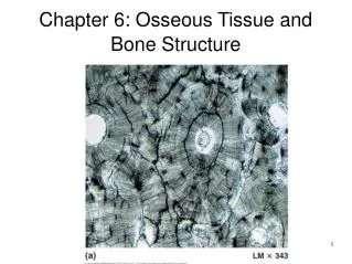
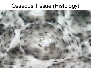
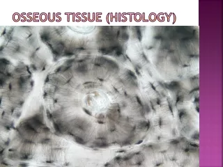
Microscopic Structure of Bone: Compact Bone Figure 6.6a, b
Microscopic Structure of Bone: Compact Bone Figure 6.6a
Microscopic Structure of Bone: Compact Bone Figure 6.6b
Microscopic Structure of Bone: Compact Bone Figure 6.6c
Spongy Bone Figure 6–6
Spongy Bone Tissue • Makes up most of the bone tissue in short, flat, and irregularly shaped bones, and the head (epiphysis) of long bones; also found in the narrow rim around the marrow cavity of the diaphysis of long bone
Spongy Bone • Does not have osteons • The matrix forms an open network of trabeculae • Trabeculae have no blood vessels
Bone Marrow • The space between trabeculae is filled with marrow which is highly vascular • Red bone marrow • supplies nutrients to osteocytes in trabeculae • forms red and white blood cells • Yellow bone marrow • yellow because it stores fat • Question: Newborns have only red marrow. Red changes into yellow marrow in some bones as we age. Why?
Location of Hematopoietic Tissue (Red Marrow) • In infants • Found in the medullary cavity and all areas of spongy bone • In adults • Found in the diploë of flat bones, and the head of the femur and humerus
Bone Membranes • Periosteum – double-layered protective membrane • Covers all bones, except parts enclosed in joint capsules (continuous w/ synovium) • Made up of: • outer, fibrous layer (tissue?) • inner, cellular layer (osteogenic layer) is composed of osteoblasts and osteoclasts • Secured to underlying bone by Sharpey’sfibers • Endosteum – delicate membrane covering internal surfaces of bone
Sharpy’s(Perforating) Fibers • Collagen fibers of the outer fibrous layer of periosteum, connect with collagen fibers in bone • Also connect with fibers of joint capsules, attached tendons, and ligaments • Tendons are “sewn” into bone via periosteum
Periosteum Figure 6–8a
Functions of Periosteum • Isolate bone from surrounding tissues • Provide a route for circulatory and nervous supply • Participate in bone growth and repair
Endosteum Figure 6–8b
Endosteum • An incomplete cellular layer: • lines the marrow cavity • coverstrabeculae of spongy bone • lines central canals • Contains osteoblasts, osteoprogenitor cells, and osteoclasts • Is active in bone growth and repair
Bone Development • Human bones grow until about age 25 • Osteogenesis: • bone formation • Ossification: • the process of replacing other tissues with bone • Osteogenesis and ossification lead to: • The formation of the bony skeleton in embryos • Bone growth until early adulthood • Bone thickness, remodeling, and repair through life
Calcification • The process of depositing calcium salts • Occurs during bone ossification and in other tissues