Download
1 / 8
80 likes | 87 Views
Characterization Techniques for Nanoparticles

E N D
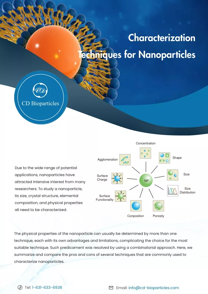
Characterization Techniques for Nanoparticles CD Concentration Shape Agglomeration Due to the wide range of potential applications, nanoparticles have attracted intensive interest from many researchers. To study a nanoparticle, its size, crystal structure, elemental composition, and physical properties all need to be characterized. Size Surface Charge NP Size Distribution Surface Functionality Conposition Porosity The physical properties of the nanoparticle can usually be determined by more than one technique, each with its own advantages and limitations, complicating the choice for the most suitable technique. Such predicament was resolved by using a combinatorial approach. Here, we summarize and compare the pros and cons of several techniques that are commonly used to characterize nanoparticles. Tel: 1-631-633-6938 Email: info@cd-bioparticles.com
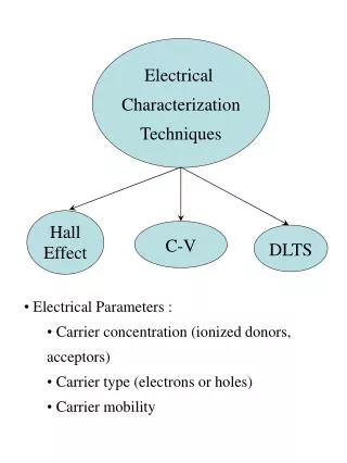
What to Measure? Shape Size Generally, nanoparticles are assumed to be spherical. While in fact, nanoparticles are in various geometric forms and irregular shapes, such as nanorods, star-shape, or cage-shape. Particle morphology can largely affect a nanoparticle’s properties, including its surface-binding capability, cellular uptake and release, and optical and plasmonic effects. High-resolution microscopy techniques can be applied to characterize the nanoparticle morphology. The size of an object is its spatial extent. For a spherical object, its size can be easily described. But to fully present the actual extension of a non-spherical object, several parameters need to be defined at the same time. Size of a macroscopic object (>1 mm) can be obtained by measuring the distance between different ends. However, for a nanoscale object, the concept of its size can convey different meanings according to the technique used to measure it, including 1) its overall physical dimensions defined by the atomic structure; 2) an effective size of the particle in a certain matrix according to its diffusion/sedimentation behavior, possibly including adsorption of matrix constituents to the nanoparticle surface, agglomeration or aggregation of the particles in the matrix; 3) an effective size of the nanoparticle, weighted by its mass/electron distribution. Porosity Porosity is usually defined as the ratio of pore volume to the total volume of a particle, and the porous structure of the nanoparticle greatly expands the range of applications. The pores on the surface of the particles drastically increase the surface-to-volume ratio, which could exceed that of non-porous particles of equal dimensions by several orders of magnitude. Moreover, porous structures contribute to the inner pore functionalization for controlled host-guest interactions, and outer functionalization for targeting purposes and drug release at the intended target site. Surface charge Zeta potential (ζ) refers to the electric potential difference between the stationary layer of charges surrounding the particles and the solution potential. It can be used to describe the surface charge and colloidal stability of nanoparticles. Generally, if the measured absolute value of zeta potential is over 15 mV, it can be considered colloidally stable. The Zeta potential depends not only on the particle itself, but also on the state of the environment, such as pH, ionic strength, and the type of ions in the suspension. Tel: 1-631-633-6938 Email: info@cd-bioparticles.com
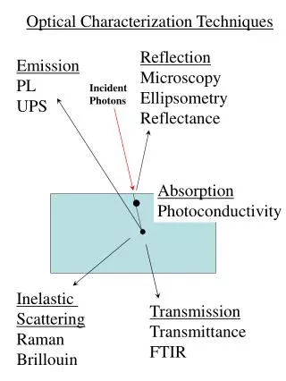
Characterization Techniques TEM Transmission electron microscopy (TEM) is a microscopy technique. In TEM, a beam of electrons is transmitted through an ultra-thin specimen and interacts with the specimen when passing through, forming an image which is then magnified and focused onto an imaging device (e.g. a fluorescent screen) or detected by a sensor (e.g. CCD camera). As a major analysis method in physical and biological sciences, TEM makes a great contribution to cancer research, virology, materials science, pollution, nanotechnology, semiconductor research, etc. Pros: Visualize particle morphology at sub-nm resolution, information on internal structure of the particles Cons: High energy beams, very expensive SEM Scanning electron microscope (SEM) is used to determine the size, shape, and morphologies of the nanoparticles. High resolution images of the surface of a sample are presented as the output. Although SEM and optical microscopes use the same principle, SEM measures the electrons (rather than photon) scattered from the sample. SEM can be used to magnify images over 200,000 times to characterize the particles. Pros: Single-particle resolution, lower energy beams than TEM, user friendly Cons: Limited penetration depth AFM Atomic force microscopy (AFM) offers ultra-high resolution in particle size measurement. It uses an atomic-scaled probe tip to physically scan samples at sub-micron level. The instrument provides a topographical map of the sample according to the forces between the tip and the sample surface. Samples can be scanned in contact or noncontact mode depending on the sample properties. Pros: High compatibility with different samples and measurement environments Cons: Samples need to be deposited on hard surface, limited throughput Tel: 1-631-633-6938 Email: info@cd-bioparticles.com
XRD X-ray diffraction (XRD) is a conventional technique to determine the crystallographic structure and morphology, of which the intensity is increased or decreased with the amount of constituent. XRD can be used to establish the metallic nature of particles and provide information on translational symmetry size and shape of the unit cell from peak positions as well as the information on electron density inside the unit cell. Pros: Rapid, provides information on crystal structure Cons: No information on particle size XAS X-ray absorption spectroscopy (XAS) has been widely used to determine the local geometric and electronic structure of the matter. This measurement is usually conducted at synchrotron radiation facilities with intense and tunable X-ray beams. Samples can be in gas, liquid, or solid phases. Pros: Highly sensitive even in very low concentrations Cons: Not a routine or readily available technique SAXS Small-angle X-ray scattering (SAXS) is a small-angle scattering technique that quantifies the nanoscale density differences in a sample. This method can determine nanoparticle size distributions, size and shape of macromolecules, pore sizes, characteristic distances of partially ordered materials, etc. Pros: High sensitivity, compatible for both dry particles and in suspension Cons: Previous knowledge of particle morphology is required for fitting the data Gas adsorption Pros: The gas adsorption is an analysis method based on the gas absorption characteristic on solid surfaces, which is usually used to measure the specific surface area and pore size distribution of materials for the size range between millimeters to nanometers. Compatible with polydisperse and aggregated samples Cons: Requires sample degassing, no information on particle morphology Tel: 1-631-633-6938 Email: info@cd-bioparticles.com
MS Mass spectrometry (MS) is an analysis method for measuring the mass-to-charge ratio (m/z) of one or more molecules in a sample. These measurements can also be used to calculate the exact molecular weight of the sample components. Moreover, for unknown compounds, they can be identified by mass spectrometers via molecular weight determination; for known compounds, they can be quantified as well as used to determine the structure and chemical properties. Pros: Information on elemental composition Cons: Sample ionization (might affect particle stability) DLS Dynamic light scattering (DLS) is used to determine the size distribution of small particles in suspension or polymers in solution. In the scope of DLS, temporal fluctuations are usually analyzed by means of the intensity or photon auto-correlation function (also known as photon correlation spectroscopy or quasi-elastic light scattering). Pros: Rapid, provides information on nanoparticle behavior in solution Cons: Highly biased toward larger particles in suspension, no information on particle shape SLS Static light scattering (SLS) measures the absolute molecular weight, utilizing the principle that builds on the relationship between the intensity of light scattered by a molecule and its molecular weight and size. In other words, larger molecules scatter higher intensity of light, that is in proportion to the molecule’s molecular weight, than smaller molecules from a given light source. Pros: Molecular weight and radius of gyration of particles in solution Cons: Highly biased toward larger particles in suspension Tel: 1-631-633-6938 Email: info@cd-bioparticles.com5
ELS Electrophoretic Light Scattering (ELS) determines the electrophoretic mobility of particles in dispersion or molecules in solution. This mobility is usually converted to Zeta potential so that materials under different experimental conditions can be compared. Pros: Rapid, typically combined with DLS Cons: Indirect estimation of zeta potential from electrophoretic mobility, ensemble-based NTA Nanoparticle tracking analysis (NTA) is used to visualize and analyze particles in liquid by relating the rate of Brownian motion to particle size. The rate of movement is only associated with the viscosity and temperature of the liquid rather than the particle density or refractive index. This method can determine the size distribution of small particles with a diameter of about 10-1000 nm in liquid suspension. Pros: Single-particle resolution, suitable for highly polydisperse samples Cons: Requires sample dilution and highly scattering particles FCS Fluorescence correlation spectroscopy (FCS) is to analyze the correlation of temporal fluctuations of the fluorescence intensity. This technique records temporal changes in the fluorescence emission intensity caused by single fluorophores passing the detection volume, by which the average number of fluorescent particles in the detection volume and their average diffusion time through the volume can be obtained. Concentration, size, and shape of the particle or viscosity of the environment can then be determined. Pros: Selectivity provided by the fluorescence detection Cons: Need of fluorescent labels (if sample is not fluorescent) Tel: 1-631-633-6938 5 Email: info@cd-bioparticles.com
AUC Analytical ultracentrifugation (AUC) can be used to characterize materials such as polymers, biopolymers, polyelectrolytes, nanoparticles, dispersions, and other colloidal systems. This technique can determine the molar mass, size, density of the particles, and interaction parameters such as virial coefficients and association constants. Pros: High-sensitivity, compatible with multimodal population Cons: High-cost equipment, highly trained users TRPS Tunable resistive pulse sensing (TRPS) is a technique that allows high-throughput single-particle measurements as colloids and/or biomolecular analytes driven through a size-tunable nanopore, one at a time. As a single-particle analyzer, it adapts the principle of resistive pulse sensing for quantitative size measurements, which monitors current flow through an aperture. Combined with the use of tunable nanopore technology, TRPS allows the passage of ionic current and particles to be regulated by adjusting the pore size. Pros: Tunable detection range, single-particle resolution, provides information on surface charge Cons: Requires (highly) conductive solutions, requires careful calibration FFF The principle of field-flow fractionation (FFF) is based on the dual effects of flow behavior and field distribution in a thin andopen channel. This method sorts and isolates nanoparticles by size or conduct size/spectroscopic characterization by uncorrelated detection methods. Pros: Highly tunable (different accumulation forces can be used), provides monodisperse sample fractions Cons: Sample recovery and choice of experimental parameters can be challenging Tel: 1-631-633-6938 Email: info@cd-bioparticles.com 5
SEC Size-exclusion chromatography (SEC) is a chromatographic method. Molecules, usually large molecules or macromolecular complexes in a solution can be separated by the differences of their size or molecular weight via SEC. Pros: Provides highly monodisperse sample fractions, compatible with industrial settings Cons: Absolute size quantification might be challenging due to particle-solid phase interaction Creative Diagnostics offers a comprehensive list of nanoparticles with precise characterization. Please visit our website for more information. References: Heera, P., & Shanmugam, S. (2015). Nanoparticle characterization and application: an overview. Int. J. Curr. Microbiol. App. Sci, 4(8), 379-386. 1. Modena, M. M., Rühle, B., Burg, T. P., & Wuttke, S. (2019). Nanoparticle Characterization: What to Measure?. Advanced Materials, 1901556. 2. Jiao, X., Tanner, E. E., Sokolov, S. V., Palgrave, R. G., Young, N. P., & Compton, R. G. (2017). Understanding nanoparticle porosity via nanoimpacts and XPS: electro-oxidation of platinum nanoparticle aggregates. Physical Chemistry Chemical Physics, 19(21), 13547-13552. 3. Mourdikoudis, S., Pallares, R. M., & Thanh, N. T. (2018). Characterization techniques for nanoparticles: Comparison and complementarity upon studying nanoparticle properties. Nanoscale, 10(27), 12871-12934. 4. Pal, S. L., Jana, U., Manna, P. K., Mohanta, G. P., & Manavalan, R. (2011). Nanoparticle: An overview of preparation and characterization. Journal of applied pharmaceutical science, 1(6), 228-234. 5. For more information, view our website: www.cd-bioparticles.com Email: info@cd-bioparticles.com Tel: 1-631-633-6938 Fax: 1-631-938-8221 Address: 45-1 Ramsey Road, Shirley, NY 11967, USA 5 5