Download
1 / 27
270 likes | 393 Views
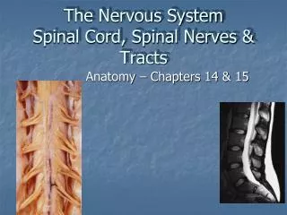
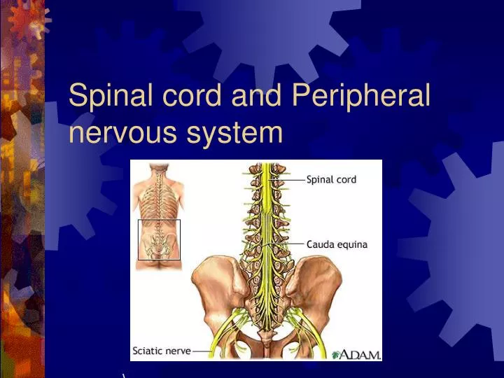
Spinal cord and Peripheral nervous system. . Spinal cord - Functions. Sensory and motor pathway. Reflex arc (spinal cord). Reflex center – Sensory receptor Sensory neuron Interneuron (association neuron) Motor neuron (effector) An effector organ. Spinal Cord Anatomy.

E N D
Spinal cord - Functions • Sensory and motor pathway
Reflex arc (spinal cord) • Reflex center – • Sensory receptor • Sensory neuron • Interneuron (association neuron) • Motor neuron (effector) • An effector organ
Spinal Cord Anatomy • Association neuron • Motor http://www.bayareapainmedical.com/wspinecrd.html • Gray Matter – “butterfly” interneurons • White Matter – myelinated
Spinal Cord tracts • Sensory • 1. Dorsal column 2. Spinothalamic • Ascending tracts • temperature, pressure, pain, light, touch
Spinal cord tracts continued Motor tracts 1. Corticospinal • Decending • Skeletal tone, voluntary muscle movement
Nerves attached to Sp. Cord • Dorsal Root Ganglia – bundle of sensory nerves • Ventral Root Ganglia – bundle of motor fibers
Somatic Nervous System • Includes all nerves in the musculoskeletal system, sense organs • Receptor (receives impulse) to Effector (muscle fiber)
Autonomic Nervous System • Motor neurons that control internal organs (involuntary) • Innervate all organs • Two divisions of
Sympathetic “Fight or flight response” Inhibits digestion Pupils dilate Accelerates heart rate Increase breathing rate. Parasympathetic Normal state Promotes digestion Pupils constrict Normal heartbeat “feed and breed” Autonomic Nervous System
The Eye: Photoreceptor • Lens – refraction and focusing • Iris – controls entrance of light into eye • Pupil – window into the eye • Choroid – blood vessels, absorbs stray light
Eye anatomy continued • Sclera – white fiborous layer, protection • Humors – • Aqueous humor – between the cornea an lens • Viterous humor – fills large cavity, gelatinous material
Eye Anatomy continued • Ciliary body – holds lens in place • Retina – contains receptors • Cones – color vision • Rods – black and white vision • Optic Nerve
Eye Anatomy Continued • Optic Nerve – picks up impulse • Ciliary muscles – controls the shape of the lens • Accommodation – • Additional focusing power • Near object – ciliary muscle contracts, lens becomes round
Physiology of sight • Focusing – light rays bent by cornea, focus on the retina, refraction and inverted
Fields of Vision Illustration Refer to Lab on eye dissection
Normal Vision 20/20 • at a distance of 20 feet, you can read a certain line (labeled 20) on the chart and that your vision is normal. • 20/40 -
Disorders of the Eye: Glaucoma – built up pressure in the eye due to lack of aqueous humor drainage