Download
1 / 43
540 likes | 1.19k Views
Spinal Cord Reflexes Peripheral Nervous System. Spinal Cord Anatomy. Extends from the foramen magnum of the skull to the first or second lumbar vertebra Provides a two-way conduction pathway from the brain to and from the brain 31 pairs of spinal nerves arise from the spinal cord

E N D
Spinal Cord Reflexes Peripheral Nervous System
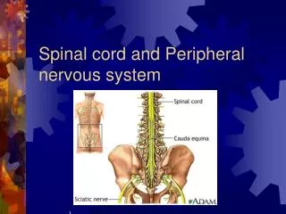
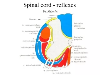
Spinal Cord Anatomy • Extends from the foramen magnum of the skull to the first or second lumbar vertebra • Provides a two-way conduction pathway from the brain to and from the brain • 31 pairs of spinal nerves arise from the spinal cord • Caudaequina is a collection of spinal nerves at the inferior end
Spinal Nerves • 31 left-right pairs of spinal nerves emerge from the cord at regular intervals (called segments). Except for the first cervical pair the spinal nerves leave the vertebral column from the intervertebral foramen between adjoining vertebrae – the first pair leaves between the skull and the first cervical vertebrae . • Cervical – 8 pairs, C1-C8 • Thoracic – 12 pairs, T1-T12 • Lumbar – 5 pairs, L1-L5 • Sacral - 5 pairs, S1-S5 • Coccygeal – 1 nerve pair
Peripheral Nervous System (PNS) Peripheral Nervous System - PNS somatic (SNS) sensory motor autonomic (ANS) sensory motor parasympathetic sympathetic
PNS: Autonomic Nervous System Motor subdivision of the PNS Consists only of motor nerves Also known as the involuntary nervous system Regulates activities of cardiac and smooth muscles and glands Two subdivisions Sympathetic division Parasympathetic division
PNS: Differences Between Somatic and Autonomic Nervous Systems
PNS: Differences Between Somatic and Autonomic Nervous Systems Central nervous system Peripheral nervous system Effector organs Acetylcholine Skeletal muscle Somatic nervous system Smooth muscle (e.g., in stomach) Norepinephrine Acetylcholine Ganglion Sympathetic division Epinephrine and norepinephrine Acetylcholine Autonomic nervous system Blood vessel Glands Adrenal medulla Acetylcholine Cardiac muscle Parasympathetic division Ganglion KEY: Postganglionic axons (sympathetic) Preganglionic axons (parasympathetic) Postganglionic axons (parasympathetic) Preganglionic axons (sympathetic) Myelination
PNS: Parasympathetic Division Preganglionic neurons originate from the craniosacral regions: The cranial nerves III, VII, IX, and X S2 through S4 regions of the spinal cord Due to site of preganglionic neuron origination, the parasympathetic division is also known as the craniosacral division Terminal ganglia are at the effector organs Neurotransmitter: acetylcholine
PNS: Sympathetic Division Preganglionic neurons originate from T1 through L2 Ganglia are at the sympathetic trunk (near the spinal cord) Short pre-ganglionic neuron and long post-ganglionic neuron transmit impulse from CNS to the effector Neurotransmitters: norepinephrine and epinephrine (effector organs)
Parasympathetic Sympathetic Eye Eye Brain stem Salivary glands Skin Cranial nerves Salivary glands Sympathetic ganglia Heart Cervical Lungs Lungs T1 Heart Stomach Thoracic Stomach Pancreas Liver and gall- bladder Pancreas L1 Liver and gall- bladder Adrenal gland Lumbar Bladder Bladder Pelvic splanchnic nerves Genitals Genitals Sacral nerves (S2 – S4)
PNS: Autonomic Functioning Sympathetic—“fight or flight” Response to unusual stimulus Takes over to increase activities Remember as the “E” division Exercise, excitement, emergency, and embarrassment • Parasympathetic—“housekeeping” activites • Conserves energy • Maintains daily necessary body functions • Remember as the “D” division • digestion, defecation, and diuresis
Peripheral Nervous System Flow to the CNS Flow out of the CNS
Peripheral Nervous System Integration occurs at many locations along the pathway. stimulus - environmental change sensation - awareness of stimulus perception - interpretation of the meaning of the stimulus
Sensory Modalities General senses: somatic and visceral Special senses smell,hearing/equilibrium taste, vision, and hearing
Classification of Sensory Receptors • Structural classification • Type of response to a stimulus • Type of stimuli they detect
Classification by Location 1. Exteroceptors 2. Interoceptors 3. Proprioceptors
Classification by Stimuli Detected • 1. Mechanoreceptors • 2. Thermoreceptors • 3. Photoreceptors • 4. Chemoreceptors • Nociceptors • Osmoreceptors
Adaptation Adaptation - generator potential or receptor potential decreases in amplitude during a maintained stimulus. Rapidly adapting - e.g. pressure, touch, smell Slowly adapting - e.g. pain, body position
Somatic Sensations Tactile touch, pressure,vibration, itch and tickle Pain fast and slow Thermal warm and cold Proprioceptive muscle spindles, tendon organs, joint receptors
Pain Sensations • Nocicceptors = pain receptors • Free nerve endings found in every tissue of body except the brain • Stimulated by excessive distension, muscle spasm & ischemia • Tissue injury releases chemicals such as kinins, or prostaglandins • Little adaptation occurs
Types of Pain • Fast Pain (acute) • occurs rapidly after stimuli (0.1 sec) • sharp pain like needle puncture or cut • not felt in deeper tissues • Slow Pain (chronic) • begins more slowly & increases in intensity • aching or throbbing pain of toothache • in superficial and deep tissues
Referred Pain • Visceral pain felt just deep to the skin overlying the stimulated organ or in a surface far from the organ • Skin area & organ are served by same segment of the spinal cord.
Pain Relief - Analgesia • Aspirin and ibuprofen block formation of prostaglandins that stimulate nociceptors • Novocaine blocks conduction of nerve impulses along pain fibers • Morphine lessens the perception of pain in the brain
Stages of Sleep Nonrapid eye movement (NREM) Rapid eye movement (REM)
Learning and Memory • Learning is the ability to acquire new information or skills through instruction or experience. • Memory is the process by which information acquired through learning is stored and retrieved. • Immediate memory- recall for a few seconds. • Short-term memory- temporary ability to recall. • Long-term memory- more permanent. • Memory consolidation.