Download
1 / 36
370 likes | 651 Views
Chapter 8: The Nervous System: The Spinal Cord and Spinal Nerves. Role of the Nervous System. Nervous system coordinates all body systems Detects and responds to stimuli Brain and spinal cord act as switching centers Nerves carry messages to and from centers. Structural Divisions.

E N D
Chapter 8:The Nervous System:The Spinal Cord andSpinal Nerves
Role of the Nervous System Nervous system coordinates all body systems • Detects and responds to stimuli • Brain and spinal cord act as switching centers • Nerves carry messages to and from centers
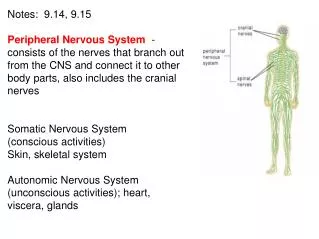
Structural Divisions • Central nervous system (CNS) • Brain • Spinal cord • Peripheral nervous system (PNS) • Cranial nerves • Spinal nerves
Functional Divisions Somatic nervous system • Controlled voluntarily • *Effectors are skeletal muscles • No further subdivisions Autonomic (or visceral) nervous system (ANS) • Controlled involuntarily • Effectors are smooth muscle, cardiac muscle, and glands • Subdivided into • Sympathetic nervous system • Parasympathetic nervous system
Overview of the Nervous System Functional Divisions of the PNS
Neurons and Their Functions Neurons • Functional cells of nervous system • Highly specialized • Unique structure
Structure of a Neuron Cell body • Nucleus • Other organelles Cell fibers • Dendrites • To cell body • Axons • Away from cell body • Some are protected by myelin sheath • Schwann cells outermost coating is neurilemma
Diagram of a motor neuron. The break in the axon denotes length. The arrows show the direction of the nerve impulse.
Types of Neurons • Sensory neurons (afferent neurons) • Conduct impulses to spinal cord, brain • Motor neurons (efferent neurons) • Conduct impulses to muscles, glands • Interneurons (central or association neurons) • Conduct information within CNS
Nerves and Tracts • Nerve: fiber bundle within PNS • *Tract: fiber bundle within CNS • Organized into fascicles • Connective tissue layers • Endoneurium • Perineurium • Epineurium
Neuroglia-astrocytes, schwann cells Neuroglia (glial cells) • Protect and nourish nervous tissue • Support nervous tissue • Aid in cell repair • Remove pathogens and impurities • Regulation composition of fluids around and between cells • Schwann cells (type of neuroglia)
The Nervous System at Work Electrical impulses sent along neuron fibers and transmitted between cells at junctions
The Nerve Impulse • Plasma membrane carries electrical charge (potential) • Plasma membrane is polarized (negative charge) • Membrane potential reverses, generates electrical charge (action potential) • Resting state • Depolarization • Na flows into cell • Repolarization • K leaves cell • Sodium/potassium (Na+/K+) pump • Myelin sheath speeds conduction
The Synapse Junction point for transmitting nerve impulse • Axon (presynaptic cell) • Dendrite (postsynaptic cell) • Synaptic cleft • Tiny gap between cells • Neurotransmitters • Epinephrine (adrenaline) • Norepinephrine (noradrenaline) • Acetylcholine • Receptors on postsynaptic cells pick up and respond to specific neurotransmitters
The Spinal Cord • Contains CSF • Links PNS and brain • Helps coordinate impulses within CNS • Contained in and protected by vertebrae
Structure of the Spinal Cord • Unmyelinated tissue (gray matter) • Dorsal horn • Ventral horn • Gray commissure • Central canal • Myelinated axons (white matter) • Posterior median sulcus • Anterior median fissure • Separates right and left portions of the anterior white matter • Ascending and descending tracts • Sensory travel ascending tract • Motor impulses travel descending tracts
The Reflex Arc • Receptor detects stimulus • Sensory neuron transmits impulses to CNS • CNS coordinates impulses and organizes response • Motor neuron carries impulses away from CNS • Effector carries out response
Typical reflex arc. Numbers show the sequence of impulses through the spinal cord (solid arrows). Contraction of the biceps brachii results in flexion of the arm at the elbow.
Reflex Activities • Simple reflex • Rapid • Uncomplicated • Automatic • Spinal reflex • Coordinated in spinal cord • Stretch reflex is example
The patellar (knee-jerk) reflex is a simple, spinal and stretch reflex
The Spinal Nerves • 31 pairs • Each nerve attached to spinal cord by two roots • Dorsal root • Dorsal root ganglion-contains sensory neurons • Ventral root • A ganglion is a collection of nerve cell bodies located outside the CNS • Nerves near end of cord travel together in the cord until each exits from its respective intervertebral foramen • Mixed nerves
Branches of the Spinal Nerves • Cervical plexus • Phrenic nerve • Brachial plexus • Radial nerve • Lumbosacral plexus • Sciatic nerve • Dermatomes
Dermatomes. A dermatome is a region of the skin supplied by a single spinal nerve.
The Autonomic NervousSystem (ANS) Regulates the action of glands, smooth muscles of hollow organs and vessels, and heart muscle • Preganglionic neuron connects spinal cord to ganglion • Postganglionic neuron connects ganglion to effector
Divisions of the Autonomic NervousSystem • Sympathetic nervous system • Parasympathetic nervous system
Sympathetic nervous system • Thoracolumbar area • Collateral ganglia • Celiac ganglion • Superior mesenteric ganglion • Inferior mesenteric ganglion • **Adrenergic system • Activated in the four E’s: excitement, emergency, embarassment, exercise
Parasympathetic nervous system • Arise in craniosacral areas • Terminal ganglia • Cholinergic system
Cellular Receptors • “Docking sites” on postsynaptic cell membranes Two types: • Cholinergic receptors • Nicotinic (bind nicotine) on skeletal muscle cells • Muscarinic (bind muscarine, a poison) on effector cells of PNS • Adrenergic receptors • Found on receptor cells of sympathetic nervous system • Bind norepinephrine, epinephrine
Functions of the Autonomic NervousSystem • Sympathetic nervous system • Fight-or-flight response • Parasympathetic nervous system • Returns body to normal • Systems generally have opposite effects on organ