Download
1 / 52
520 likes | 527 Views
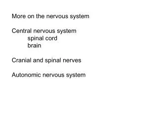
1. Nervous system generalities, spinal cord. Dr. Phd. Bartoș Dana. Nervous system. General features Ganglion: a collection of cell bodies located outside the Central Nervous System Nucleus : a collection of cell bodies located inside the Central Nervous System. Nervous system.

E N D
1. Nervous system generalities, spinal cord Dr. Phd. Bartoș Dana
Nervous system • General features • Ganglion: • a collection of cell bodies located outsidethe Central NervousSystem • Nucleus: • a collection of cellbodieslocatedinsidetheCentral NervousSystem
Nervous system • General features • Nerve • a group of fibers (axons) outside the CNS • spinal nerves contain the fibers of the sensory and motor neurons • does not contain cell bodies • Tract (fascicle) • a group of fibers (axons) inside the CNS • spinal tracts carry information up or down the spinal cord, to or from the brain • are always part of white matter
Nervous system Gray matter White matter • Area of unmyelinated neurons where cell bodies and synapses occur • In the spinal cord the synapses between sensory and motor and interneurons occurs • cell bodies of the interneurons and motor neurons • an area of myelinated fiber tracts
Neuron Structure • have the ability to gather and transmit electrochemical signals • Formed by: body, axon, dendrites • Cell body: if the cell body dies, the neuron dies • Axon - long, cable-like projection of the cell -carries the electrochemical message (nerve impulse or action potential) along the length of the cell. -depending upon the type of neuron, axons can be covered with a thin layer of myelin, like an insulated electrical wire. Myelin is made of fat, and it helps to speed transmission of a nerve impulse down a long axon -myelinated neurons are typically found in the peripheral nerves (sensory and motor neurons), while non-myelinated neurons are found in the brain and spinal cord.
Neuron Structure • Dendrites or nerve endings - small, branch-like projections of the cell make connections to other cells and allow the neuron to talk with other cells or perceive the environment. -they can be located on one or both ends of the cell.
Basic Neuron Types • many sizes • Some types of neurons:- Bipolar (Interneuron)- Unipolar (Sensory Neuron)- Multipolar (Motoneuron)- Cortical Pyramidal Cell
Basic Neuron Types • Neurons also vary with respect to their functions: • Sensory neurons carry signals from the outer parts of your body (periphery) into the central nervous system • Motor neurons (motoneurons) carry signals from the central nervous system to the outer parts (muscles, skin, glands) of your body • Receptors sense the environment (chemicals, light, sound, touch) and encode this information into electrochemical messages that are transmitted by sensory neurons • Interneurons connect various neurons within the brain and spinal cord
Meninges • Connective tissue membranes • Dura mater: outermost layer; continuous with epineurium of the spinal nerves • Arachnoid mater: thin and wispy • Pia mater: bound tightly to surface • Forms the filum terminale • anchors spinal cord to coccyx • Forms the denticulate ligaments that attach the spinal cord to the dura
Meninges • Spaces • Epidural: external to the dura • Anesthestics injected here • Fat-fill • Subdural space: serous fluid • Subarachnoid: between pia and arachnoid • Filled with CSF
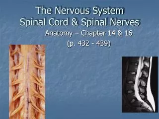
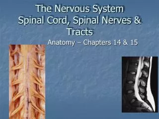
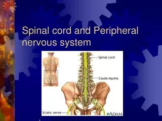
Spinal Cord • Runs through the vertebral canal • Extends from foramen magnum to second lumbar vertebra • Regions • Cervical • Thoracic • Lumbar • Sacral • Coccygeal • Gives rise to 31 pairs of spinal nerves • All are mixed nerves
Spinal Cord • Not uniform in diameter • Cervical enlargement: supplies upper limbs • Lumbar enlargement: supplies lower limbs • Conus medullaris- tapered inferior end • Ends between L1 and L2 • Cauda equina - origin of spinal nerves extending inferiorly from conus medullaris.
Inferior End of Spinal Cord • Conus medullaris - inferior end of spinal cord proper • Cauda equina - individual spinal nerves within spinal canal • Filum terminale - filamentous end of meninges, "tie-down"
SPINAL NERVE mixed nerve, which carries motor, sensory, and autonomic signals between the spinal cord and the body 31 pairs formed from the combination of nerve fibers from its dorsal and ventral roots dorsal root is the afferent sensory root ventral root is the efferent motor root
SPINAL NERVE Outside the vertebral column, the nerve divides into branches Dorsal Ventral Communicants White Gray
SPINAL NERVE The dorsal ramus contains nerves that serve the posterior portions of the trunk carrying visceral motor, somatic motor, and somatic sensory information to and from the skin and muscles of the back
SPINAL NERVE The ventral ramus contains nerves that serve the remaining anterior parts of the trunk and the upper and lower limbs carrying visceral motor, somatic motor, and sensory information to and from the ventrolateral body surface, structures in the body wall, and the limbs
SPINAL NERVE The rami communicantes contain autonomic nerves that serve visceral functions carrying visceral motor and sensory information to and from the visceral organs
1. posterior horn 2. anterior horn 3. intermediate zone 4. lateral horn 5. posterior funiculus 6. anterior funiculus 7. lateral funiculus 8. Lissauer's tract 9. anterior medianfissure 10. posterior median sulcus 11. anterolateral sulcus 12. posterolateralsulcus 13. Posteriorintermediate sulcus
Cross Section of Spinal Cord • Anterior median fissure and posterior median sulcus • deep clefts partially separating left and right halves • Gray matter: neuron cell bodies, dendrites, axons • Divided into horns • Posterior (dorsal) horn • Anterior (ventral) horn • Lateral horn • White matter • Myelinated axons • Divided into three columns • Ventral • Dorsal • lateral • Each of these divided into sensory or motor tracts
Cross section of Spinal Cord • Commissures: connections between left and right halves • Gray with central canal in the center • White • Roots • Spinal nerves arise as rootlets then combine to form dorsal and ventral roots • Dorsal and ventral roots merge laterally and form the spinal nerve
White Matter in the Spinal Cord • Divided into three columns – posterior, lateral, and anterior • Columns contain 3 different types of fibers (ascending, descending, transvers) • Fibers run in three directions • Ascending fibers - compose the sensory tracts • Descending fibers - compose the motor tracts • Commissural (transverse) fibers - connect opposite sides of cord
Organization of Spinal Cord Gray Matter • Recall, it is divided into horns • Dorsal, lateral (only in thoracic region), and ventral • Dorsal half – sensory roots and ganglia • Ventral half – motor roots • Based on the type of neurons/cell bodies located in each horn, it is specialized further into 4 regions • Somatic sensory (SS) - axons of somatic sensory neurons • Visceral sensory (VS) - neurons of visceral sensory neur. • Visceral motor (VM) - cell bodies of visceral motor neurons • Somatic motor (SM) - cell bodies of somatic motor neurons
Gray Matter: Organization Figure 12.31
Neurons of grey matter • Golgi I (long axon) • Golgi II (short axon)
GOLGI I • 3 types 1. Anterior radicular neurons • Form the ant. root of the spinal nerve • 4 types: 1. α and β (somatomotor) • receive aferences from pyramidal, extrapyramidal fascicles and spinal ggl. • axons end on the motor plates of striate muscles • before leaving the gray matter they give up a collateral to synapse with Renshaw neurons • the Renshaw neurons receive an excitatory collateral from the alpha neuron's axon after that they will send an inhibitory axon to synapse with the cell body of the initial alpha neuron and/or an alpha motor neuron of the same motor pool
Anterior radicular neurons 2. γ - takes part in the process of muscle contraction - receive afferences from reticulospinal fascicle - axons will go on the neuromuscular bundles - do not directly adjust the lengthening or shortening of muscles - their role is to keep the muscle spindle taut - these neurons play a role in adjusting the sensitivity of muscle spindles
Anterior radicular neurons 3. Lissomotors (Sympathetic visceromotor) - mostly at the level of C2-L2 - receive afferences from hypothalamus, reticular substance, periphery - at the level of lateral horns - their axons arrive in the trunk of the spinal nerve white communicating branch sympathetic laterovertebral ggl. chain (preganglionic fibers) - some synapse here postganglionic fibers through splanchnic nerves organs - some don’t synapse go through splanchnic nerves synapse in previsceral ggl. postganglionic fibers organs
Anterior radicular neurons 4. Parasympathetic visceromotors - mostly at the level S2-S4 - axons are preggl. fibers that will synapse in the previsceral or intramural ggl. sacral parasympathetic fibers splanchnic pelvic nerves hypogastric ggl.
GOLGI I 2. Posterior radicular neurons - not all the anatomists agree with them - supposed to by located at the root of anterior and posterior horns - supposed to be parasympathetic neurons
GOLGI I 3. Coordinal neurons - mostly on the posterior horns - afferences come through posterior root of the spinal nerve from the spinal ggl. - efferences go through white matter expecially lateral and anterior colums - they can have a homolateral, heterolateral and bylateral path
GOLGI II • Short axon • Do not leave the gray substance • Afferent fibers from the descending tracts • Synapse with anterior homo or heterolateral radicular neurons
Laminae I–IVcorrespond to the dorsal part of the dorsal horn • Laminae V-VIlie at the base of the dorsal horn • Laminae VIIcorresponds to the intermediate area between dorsal horn and lateral horn lamina I corresponds approximately to the marginal substance lamina II corresponds approximately to the substantia gelatinosa, is characteristically distinguished from adjacent laminae by he almost total lack of myelinated fibres laminae III and IV corresponds approximately to the nucleus proprius laminae V and VI corresponds approximately to the Bechterew nucleus lamina VII within the thoracic cord includes the lateral horn, it contains three important nuclear groups: nucleus thoracicus posterior or Clarke’s column, intermediolateral nucleus, intermediomedial nucleus
Laminae VII • corresponds to the intermediate area between dorsal horn and anterior horn • within the thoracic cord includes the lateral horn and not the anterior • in the cervical and lumbar enlargements it extends laterally and ventrally troughout the ventral horn • lamina VII • contains three important nuclear groups: nucleus thoracicus posterior or Clarke’s column, intermediolateral nucleus, intermediomedial nucleus
Laminae VIII • corresponds to the base of the ventral horn in the thoracic region • is restricted to the medial aspect of the ventral horn in the cervical and lumbar enlargements • Laminae IX • in the thoracic cord, the nuclear cell groups are embedded within lamina VIII • in the cervical and lumbar enlargements, the nuclear cell groups are embedded within both lamina VII and lamina VIII • Lamina X • surrounds the central canal and consists of the dorsal and ventral grey commissures
Second neuron for the exteroceptive andproprioceptivepathway Waldeyer Rolando Of the Head/ Waldeyer Clarke Bechterew
10. C8-L2 C8-L2- vasoconstrictors C8-T3- dilatator of the pupil T2-T3- cardio accelerator T2-T4- viscera of the posterior mediastinum T5-T10- viscera of the abdomen T3-L1- inhibitors of the bowel movements T12-L2- pelvic viscera T1-L2 Only at the level of intumescences Only at the level of intumescences Accesory nucleus (C1-C4) C1-S4 (C2-T2 and L2-L4) 4. colonne en torsade of Laruelle (L5-S5)
Arterial Supply • - Spinal Arteries Anterior (1) & Posterior (2) Spinal Artery from Vertebral artery • - Radicular Arteries ----- Segmental arteries from Vertebral, Ascending Cervical, Intercostal andLumbar Artery