Download
1 / 7
70 likes | 246 Views
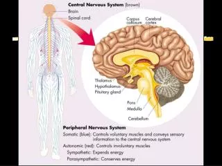
Peripheral Nervous System. General Organization. Cranial Nerves. Spinal Nerves. Reflex Activity. Autonomic Nervous System. Somatic Sensory System. Home. Exit. BASIM ZWAIN LECTURE NOTES. General Organization. Motor endings. Background. Sensory receptors. Nerves.

E N D
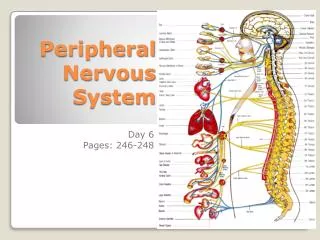
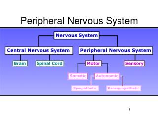
Peripheral Nervous System General Organization Cranial Nerves Spinal Nerves Reflex Activity Autonomic Nervous System Somatic Sensory System Home Exit BASIM ZWAIN LECTURE NOTES
General Organization Motor endings Background Sensory receptors Nerves Nature of stimulus detected a. Mechanoreceptors i. Touch, vibration, pressure, stretch b. Thermoreceptors i. Temperature changes c. Photoreceptors i. Light energy ii. Exclusively in the retina d. Chemoreceptors i. Chemical in solution e. Nociceptors i. Pain Generalized sensory receptors Types a. Neuromuscular junction i. Contact between motor neuron and muscle ii. Release ACh b. Varicosities i. Contact between autonomic motor Endings and visceral effectors and organs, Smooth and cardiac muscle Location a. Exteroceptors i. Surface of skin b. Interoceptors i. Visceroceptors ii. Visceral organs and blood vessels c. Proprioceptors i. Musculoskeletal organs Classification based on nature of information a. Sensory (afferent) nerves i. Sensory information from periphery to CNS b. Motor (efferent) nerves i. Motor information from CNS to periphery c. Mixed nerves i. Include sensory and motor 1. Function a. Connect brain with outside world i. CNS function is dependent on information 2. Structural components a. Sensory receptors b. Peripheral nerves and ganglia c. Efferent motor endings Complexity a. Simple i. Most sensory receptors (generalized) b. Complex i. Special senses (vision, audition, olfaction, gustation) b. Encapsulated i. Meisner’s corpuscles—low frequency vibration) ii. Pacinian corpuscles—high frequency iii. Ruffini’s corpuscles—deep pressure iv. Muscle spindles—muscle stretch v. Golgi tendon organs—tendon stretch Classification based on site of origin a. Cranial nerves i. Brain origin b. Spinal nerves i. Arise from spinal cord Parallel bundles of peripheral axons a. Enclosed by connective tissue b. Some may be myelinated Function a. Activate effectors i. Release of neurotransmitter a. Free dendritic endings (unencapsulated) i. Free ii. Merkel discs iii. Root hair plexus Home Exit BASIM ZWAIN LECTURE NOTES
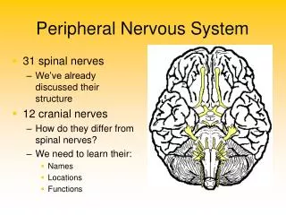
Cranial Nerves Home Exit BASIM ZWAIN LECTURE NOTES
Spinal Nerves Nomenclature Structure Nerves plexuses Dermatomes 1. Dorsal and ventral rootlets 2. Dorsal and ventral root 3. Dorsal root ganglion 4. Spinal nerve 5. Dorsal ramus of spinal nerve 6. Ventral ramus of spinal nerve 7. Rami communicantes a. Autonomic fibers 8. Sympathetic chain ganglion 3. Fibers of different ventral rami cross and are redistributed a.Branches contain fibers originating from different spinal nerves b.Innervation arrives via multiple routes i. More than a single spinal nerve serves each limb muscle Named for the level of the vertebral column from which the nerves exits a. 31 spinal nerves i. 8 cervical (C1 – C8) ii. 12 Thoracic (T1 – T8) iii. 5 Lumbar (L1 – L8) iv. 5 Sacral (S1 – S8) v. 1 Coccygeal (C0) 1. Specific to ventral rami 2. Types a. Cervical b. Brachial c. Lumbar d. Sacral regions • Area of skin innervated by the • cutaneous branch of a single • spinal nerve • 2. All spinal nerves (except C1) • Have dermatomes • 3. Dermatomes overlap Home Exit BASIM ZWAIN LECTURE NOTES
Reflex Activity Background Components of a reflex arc Stretch and deep tendon reflexes Muscle spindles 1. Receptor (site of stimulus action) 2. Sensory neuron (transmits the afferent impulse to the CNS) 3. Integration center a. Monosynaptic reflex (single synapse) b. Polysynaptic (multiple synapses with chains of interneurons) 4. Motor neuron (conducts efferent impulse from integration center to effector 5. Effector (muscle fiber or gland) Sequence of events a. Stretching muscle activates muscle spindle b. Impulse carried by primary sensory fiber to spinal cord c. Activates alpha motor neuron i. Sends efferent signal to muscle (effect) d. Stretched muscle contracts e. Antagonist muscle is reciprocally inhibited 1. Stimulus-response sequence a. Unlearned b. Unpremeditated c. Involuntary 2. Mediated by spinal cord circuits a. Information may ultimately relayed to the brain a. Consist of intrafusal fibers b. Wrapped by afferent sensory endings i. Type Ia fibers ii. Type II fibers c. Gamma (g) efferent fibers i. Innervate contractile region of spindle ii. Maintain spindle sensitivity Extrafusal muscle fibers a. Skeletal muscle b. Innervated by alpha (a) motor neurons Home Exit BASIM ZWAIN LECTURE NOTES
Autonomic Nervous System 10. Parasympathetic response 9. Sympathetic response 5. Neurotransmitter a. ACh acts locally i. ACh always has a stimulatory effect b. NE has spreads far and can exert its effects over long distances when circulated in the blood c. Adrenergic receptors i. Alpha—stimulatory ii. Beta—inhibitory (except in the heart when it is excitatory) 8. Function a. Divisions work in concert b. Parasympathetic dision i. Maintenance of function ii. Energy conservation c. Sympathetic division i. Emergence ii. Intense muscular activity 2.Cell bodies of all lower autonomic motor neurons lie outside the CNS a. Autonomic ganglia b. Neurons are postganglionic c. Driven by preganglionic neurons whose cell bodies are in the spinal cord or brainstem 6. Fiber length a. Parasympathetic i. Long preganglionic ii. Short postganglionic b. Sympathetic i. Short preganglionic ii. Long postganglionic Somatic Nervous System 1. Voluntary a. Voluntary muscle movement 2. Sensory information to the CNS 3. Organization of cell bodies a. Lie within spinal cord or brainstem b. Targets are controlled monosynaptically a. Pupil dilated b. Secretory responses inhibited c. Stimulates sweating d. Heart function i. Increases rate ii. Dilates coronary vessels a. Pupils constrict b. Stimulates secretory activity i. Salivation c. Heart function i. Decreases rate ii. Constricts coronary vessels 4. Divisions differ based on: a. Neurotransmitter type b. Fiber length c. Location of ganglia d. Function i. Increase metabolic rate i. Glucose is released into blood ii. Lipolysis j. Increased alertness h. Causes ejaculation (vaginal reverse peristalsis) e. Increased blood pressure i. Constricts most vessels f. Bronchioles dilate g. Decreased activity of digestive system h. Piloerection 3. Divisions a. Sympathetic b. Parasympathetic d. Constricts bronchioles e. Increases activity of digestive system f. Causes erection (penis and clitoris) i. Vasodilation 1.Autonomic Nervous System is involuntary (autonomic functions are carried out without conscious, voluntary control) Home Exit BASIM ZWAIN LECTURE NOTES
Somatic Sensory System Mechanical Senses Somatic Sensory Pathways Somatosensory Cortex Organization of Somatic Sensory Information Pain and Its Control Background Types of receptors Types of somatic sensation receptors 1. Mechanoreceptors--sensitive to physical distortion 2. Nociceptors--respond to damaging stimuli 3. Thermoreceptors--sensitive to changes in temperature 4. Proprioceptors--monitor body position 5. Chemoreceptors--respond to certain chemicals DCML Pathway 1. In the DCML pathway information ascends through the dorsal column on the ipsilateral side of the spinal cord 2. Synapses in the medulla 3. Crosses over and ascends via the medial lemniscus to the thalamus (VP) 4. Synapses in VP thalamus 5. Projects to the cortex Spinal segments 1. 30 spinal segments consisting of paired dorsal and ventral roots 2. Spinal segments are divided into 4 groups: cervical, thoracic, lumbar, sacral 3. Each segment is named after vertebra from which nerves emerge (cervical: C1 - C8, thoracic: T1 - T12, lumbar: L1 - L5 & sacral: S1 - S5) c. Two different mechanosensitive proprioceptors: i. Muscle spindles-consist of specialized intrafusal muscle fibers distributed among ordinary (extrafusal) muscle fibers; detect changes in muscle length ii. Golgi tendon organs-distributed among collagen fibers in tendons and detects changes in muscle tension Somatotopy 1. Mapping of the body's surface sensations onto a brain structure 2. Features of the map: a. Not continuous, b. Not scaled to the human body, c. Relative size of the cortex devoted to each body part is correlated with the density of sensory input (i.e., lips versus the skin on your calf) & d. Size is related to importance of the sensory input (i.e., finger tip versus elbow) • 1. Mechanoreceptors • Pacinian (sensitive to vibration (250-350 Hz), • involved in fine discrimination of texture or • other moving stimuli that cause vibrations • b. Meissner's corpuscle (most common • receptor in glabrous skin (smooth, hairless), • sensitive to vibration (low frequency, 30-50 Hz) Experiment--three beakers of water: one cold, one hot, one lukewarm. One finger from one hand into hot; one finger from the other hand into cold. After some time period, immerse both simultaneously into the lukewarm. The finger from the hot senses the water to be cold and the finger from the cold senses the same water to be hot. Why? Adaptation--the hot and cold receptors adapted (stopped firing). When immersed in lukewarm, only the unadapted receptors were available. You need both to sense lukewarm, etc. 4. Proprioceptors a. Body position i. Where the body is ii. Direction of movement iii. Speed of movement b. Receptors in the skeletal muscles (more in movement lecture) 2. Nociceptors a. Free, unmyelinated nerve endings b. Signal that body tissue is being damaged c. In most tissues, not brain d. Types of damage detected i. Mechanical--strong pressure (sharp objects) ii. Thermal (different from temperature)-- Active when tissues begin to be destroyed iii. Chemical--environmental agents or those from tissues itself--pH, histamine, etc. Characteristics 1. Pain is influenced cognitively 2. Hyperalgesia a. Tissue already damaged is much more sensitive to pain i. Nociceptors are sensitized by various substances released by damaged tissue (protaglandins, histamines, etc.) 3. Thermoreceptors a. Brain temperature is tightly regulated (close to 37C, brain function changes above and below that temperature) b. Specialized receptors that can perceive changes in temperature as small as 0.01C. c. Two types (warm--begin firing at 30C-45C (above causes damage and pain) & cold-- below 35C to 10C) Information carried in each pathway remains separate 1. Segregated all the way to the cortex 2. Thalamus a. Ventral posterior (VP) nucleus receives the information and projects to the somatosensory cortex Mechanism of function 1. Stimuli applied to skin deform or change receptor a. Alters the ionic permeability of the receptor creating generator potentials i. Trigger action potentials 3. Brain chemicals a. Endorphins i. Share many opioid properties and bind to opioid receptors in the brain ii. Opioid receptors are throughout body, but especially in brain and particularly in brain areas that process and modulate nociceptive information (PA, raphe, and spinal cord) Classification 1. Free nerve endings a. Nociceptors b. Thermoreceptors 2. Encapsulated a. Most cutaneous receptors Pathways 1. Dorsal column-medial lemniscal pathway DCML a. Touch and proprioception 2. Spinothalamic pathway ST a. Pain and temperature Posterior parietal lobe 1. Primary somatosensory cortex receives simple segregated streams of sensory information 2. Integration takes place in the posterior parietal cortex 2. Several brain regions can suppress pain a. PAG (periacqueductal gray matter) project to the raphe (serotonin) that sends axons to the spinal cord (5-HT is inhibitory, block synaptic activity) ST Pathway 1. Information crosses to the contralateral side in the spinal cord 2. Ascends via the spinothalamic tract 3. Synapses in the thalamus (VP) 4. Projects to the cortex. 2. Responds to many kinds of stimuli (usually mechanical) 3. At least four senses (temperature, body position, touch & pain) 4. Place, pressure, sharpness, texture, and duration can be accurately gauges Differences between somatic senses and other senses 1. Receptors are distributed throughout the body as opposed to being concentrated at small, specialized locations Regulation of pain 1. Pain can be modified by non-painful sensory input (i.e., rub the skin around a bruise) a. Gate Theory of Pain-circuit in spinal cord dorsal root 3. Characteristics a. Overlap between the dermatomes b. Cervical dermatomes: above the sternum c.Thoracic dermatomes: top of sternum to waist d.Lumbar dermatomes: front of legs &stomach e.Sacral dermatomes (back of legs & genitals) • Mechanical energy • 1. Easily differentiated • Stimulus frequency • b. Stimulus pressure • c. Receptive field Dermatomes 1. Segmental organization of spinal nerves & sensory innervation of skin are related 2. Area of skin innervated by the dorsal roots of a single spinal segment is a dermatome c. Ruffini's ending-not well understood d. Mercel's disks (light pressure and Tactile discrimination e. Hair follicle receptor Anatomy 1. Parietal lobe a. Post-central gyrus i. Most complex processing occurs in cortex Note: Like other sensory receptors, temperature receptors adapt. They respond to sudden changes in temperature. Two basic systems 1. Pain and temperature 2. Touch and Proprioception Nociception 1. Sensory process that provides signals that trigger pain Home Exit BASIM ZWAIN LECTURE NOTES