Download
1 / 42
650 likes | 1.59k Views
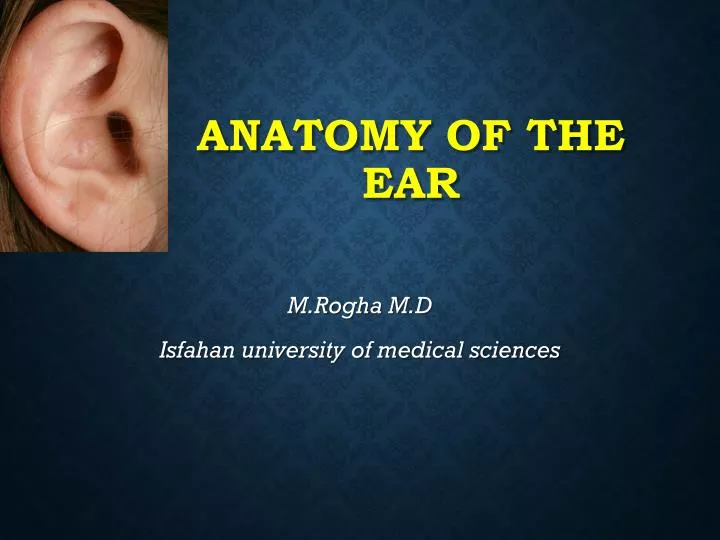
Anatomy of the Ear. M.Rogha M.D Isfahan university of medical sciences. Major Divisions of the Ear. Peripheral Mechanism. Central Mechanism. External ear , Auris externa Auricula Meatus acusticus externus Middle ear , Auris media Cavitas tympani Membrana tympanica Ossicula auditus

E N D
Anatomy of the Ear M.Rogha M.D Isfahan university of medical sciences
Major Divisions of the Ear Peripheral Mechanism Central Mechanism
External ear, Auris externa Auricula Meatus acusticus externus Middle ear, Auris media Cavitas tympani Membrana tympanica Ossicula auditus Tuba auditiva Inner ear, Auris interna Labyrinthus membranaceus - Labyrinthus vestibularis - Labyrinthus cochlearis Labyrinthus osseus - Vestibulum - Canales semicirculares ossei - Cochlea - Meatus acusticus internus
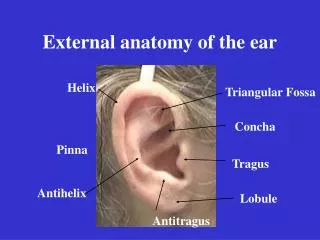
Outer Ear Pinna Preauricular Tags Preauricular Pits EAM Cerumen Pinna External Auditory Meatus Function EAM resonance
Function of Outer Ear • Collects sound • Localization • Resonator • Protection
Pinna • The visible portion that is commonly referred to as "the ear" • Helps localize sound sources • Directs sound into the ear • Each individual's pinna creates a distinctive imprint on the acoustic wave traveling into the auditory canal
External Auditory Meatus • Extends from the pinna to the tympanic membrane • About 26 mm in length and 7 mm in diameter in adult ear. • Size and shape vary among individuals. • Protects the eardrum • Resonator • Provides about 10 decibels (dB) of gain to the eardrum at around 3,300 Hertz (Hz). • The net effect of the head, pinna, and ear canal is that sounds in the 2,000 to 4,000 Hz region are amplified by 10 to 15 dB. • Sensitivity to sounds greatest in this frequency region • Noises in this range are the most hazardous to hearing
Outer ear Tissues: elastic cartilage covered with skin A.Meatus acusticus externus besides the hair follicles and fat glands contains: Glandulaeceruminosae – modified sweat glands on the lateral wall of the canal. Сerumеn (ear wax) combination of wax andfat glands secret and desquamated epithelial cells.
Middle Ear Tympanic Cavity Tympanic Membrane Ossicles Middle Ear Muscles Eustachian Tube Mastoid
Function of Middle Ear • Conduction • Conduct sound from the outer ear to the inner ear • Protection • Creates a barrier that protects the middle and inner areas from foreign objects • Middle ear muscles may provide protection from loud sounds • Transducer • Converts acoustic energy to mechanical energy • Converts mechanical energy to hydraulic energy • Amplifier • Transformer action of the middle ear • only about 1/1000 of the acoustic energy in air would be transmitted to the inner-ear fluids (about 30 dB hearing loss)
Tympanic cavity • Volume – 1.5 ml • Form – flatten drum • Structure – six walls: • - Lateral • - Medial • - Anterior • - Posterior • - Superior • -Inferior
Tympanic Membrane • Separates outer ear from middle ear • Barrier from foreign objects • Cone-shaped in appearance • about 17.5 mm in diameter • Vibrates in response to sound waves. • The membrane movement is incredibly small • as little as one-billionth of a centimeter
Tympanic membrane Two parts: Pars flaccida – upper, thin, loose Pars tensa – lower, tense Three layers: 1. Outer, cutaneous – continuation of the canal skin. No hairs and glands. 2. Middle, fibrous – elastic fibers. 3. Inner, mucous – tympanic cavity lining
Medial wall, paries labyrinthicus Most complex. On this wall are distinguished: -fenestra vestibuli -fenestra cochleae -promontorium -prominentia canalis semicircularis lateralis - prominentia canalis facialis Superior wall, paries tegmentalis Separates tympanic from cranial cavity. Children less than 2 years– infections of the middle ear can pass to the cranial cavity.
Inferior wall, paries jugularis Separates tympanic cavity fromfossa jugularis Anterior wall, paries caroticus Separates tympanic cavity fromcanalis caroticus • canalis musculotubularis Posterior wall, paries mastoideus Composed of: • Styloid complex ofProcter • Antrum mastoideum • Fossa incudis
Auditory (Eustachian) tube • Connects tympanic cavity with pharynx • Two openings: • ostium pharyngeum tubae • ostium tympanicum tubae. • Two parts: • bony • cartilagenous • Function: • Equalizes pressure on both sides of tympanic membrane for optimal hearing.
Ossicles • Malleus (hammer) • Incus (anvil) • Stapes (stirrup) smallest bone of the body
Inner Ear Vestibular semicircular canals utricle and saccule Cochlear traveling wave traveling wave traveling wave pathologies Auditory Vestibular
Inner ear Two compartments: (а) Bony labyrinth and (b) Membraneous labyrinth. Bony labyrinth: complex cavity in dense bone (pars petrosa) Parts of the bony labyrinth: a.Vestibulum. b. Semicircular canals. c.Cochlea.
Bony labyrinth. Labyrinthus osseus Vestibulum and semicircular canals
Vestibulum Two walls: Externalandinternal. External wall has • Fenestra vestibuli. Internal wall has: • Recessus ellipticus • Recessus sphericus • Recessus cochlearis • Maculae cribrosae superior, medius, inferior
Openings into vestibulum a. Fenestra vestibuli. b. Fenestra cochleae. c. Openings (5) of the semicircular canals d. Aqueductus vestibuli
Semicircular canals 3: anterior, posterior and lateral. Have ampulla and crus. Canalis semicircularis lateralis –horizontal. - eminentia canalis semicircularis lateralis on the medial wall of tympanic cavity. Canalis semicircularis anterior –frontal. -eminentia arcuataonpars petrosa ofos temporale. Canalis semicircularis posterior –sagittal
Labyrinthus osseus. Cochlea Meatus acusticus internus Cochlea
Cochlea • Cone-shaped: baseandapex. • Canalis spiralis cochleae -promontorium, on the medial wall of tympanic cavity. • Modiolus -canales longitudinales modioli. • Lamina spiralis ossea • hamulus Dividescanalis spiralis cochleaeinto: • Scala tympani • Scala vestibuli
Function of Inner Ear • Converts mechanical sound waves to neural impulses that can be recognized by the brain for: • Hearing • Balance
Membraneous labyrinth.Labyrinthus membranaceus • Closed system of sacs and ductsunderling the bony labyrinth. • Filled with endolymph. • Two parts: vestibular& cochlear.
Vestibular labyrinth Composed of : • Two bags - sacculus et utriculus • Three ductus semicirculares • Oneductus endolymphaticus.
Balance • Linear motion • Rotary motion
Macula utriculi (sacculi) Otoliths. Statoconia
Static balance Maculae react to gravitational forcesand participate in maintaining the static balance.
Dynamic balance Cristae ampullares react to rotatory movementsand paticipate in dynamic balance.
Cochlea • The cochlea resembles a snail shell and spirals for about 2 3/4 turns around a bony column • Within the cochlea are three canals: • Scala Vestibuli • Scala Tympani • Scala Media
Cochlear labyrinth Spiral canal -ductus cochlearis. Occupiesscala mediaofthe spiral canal. Has two blind ends - cecum vestibulare and cecum cupulare. Has three walls: • paries vestibularis • paries externus • paries tympanicus- organ of Corti, basal membrane
Organ of Corti • End organ of hearing