Download
1 / 42
430 likes | 506 Views
Slides prepared and compiled by highly experienced ENT teacher, Dr. Krishna Koirala from Nepal, for teaching undergraduate and postgraduate ENT students in the field of otorhinolaryngology. <br>A clear and concise explanation of the basic concepts in the subject matter concerned. <br>He is the Head of department with a sound knowledge in the field of ENT to teach both undergraduate and postgraduate ENT students

E N D
Anatomy of ear and mastoid Dr. Krishna Koirala
Paired sensory organs comprising of • Auditory system involved in the detection of sound • Vestibular system involved in maintaining body balance and equilibrium • Divided anatomically and functionally into • External ear • Middle ear • Inner ear • All three regions are involved in hearing • Inner ear is involved in body balance and equilibrium
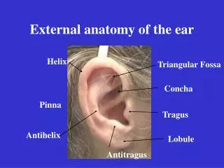
Pinna • Framework formed by yellow elastic cartilage except in the lobule and incissura terminalis • Functions • Collect and direct sound waves through the ear canal to the tympanic membrane • Protect the tympanic membrane • Importance : Graft material for middle ear & other reconstructive surgeries
Helix: Slightly curved rim of the auricle • Antihelix: Broader curved eminence anterior to helix • Concha : Deep cavity in front of the helix • Cymba conchae : Depression between the antitragus and ascending crus of the helix (surface landmark of mastoid antrum) • Tragus • Lobule:Structure made up of areolar tissue & fat without cartilage
Sensory Nerve supply of pinna • Lateral surface • Upper 2/3 : Auriculotemporal nerve (cranial nerve V) • Lower 1/3 : Greater auricular nerve (C2,3) • Medial Surface • Upper 1/3: Lesser occipital nerve (C2) • Lower 2/3 : Greater auricular nerve (C2, 3) • Posterior concha and antihelix : Auricular b/o Vagus • Facial : Small region at the root of concha
Extends from bottom of concha to the tympanic Membrane • 24 mm long in adults • Lateral 1/3 (8 mm) : Cartilaginous, directed upwards, backward and medially • Medial 2/3 (16 mm) : Bony, directed downwards, forward and medially • Pinna to be pulled upwards, backwards and laterally to straighten the external auditory canal in adults
Only cartilaginous skin has hair follicles, ceruminous and pilosebaceous glands (wax) • Cartilaginous fissure of Santorini and bony foramen of Huschke present in anterior wall infection / metastasis to and from the parotid gland
Middle ear cavity Epitympanum Mesotympanum Hypotympanum Protympanum Post- tympanum Middle ear cleft • Contents • Middle ear cavity • Attic ,aditus, antrum • Mastoid air cell system • Eustachian tube • Develops from tubo -tympanic recess
AD Ant ATTIC ME ET
Contents of middle ear • 3 Ossicles : malleus, incus, stapes • 2 Nerves : Chorda tympani, Tympanic plexus • 2 Muscles: Tensor tympani, stapedius • Air • Mucosal folds & ligaments • Blood vessels
Tympanic Membrane • Partition between the external and middle ear • Obliquely set with 550 to floor • Dimension: 10 mm x 8 mm x 0.1 mm • Parts • Pars Tensa • Pars Flaccida (Shrapnel's membrane)
PF PT
Landmarks of TM • Lateral process of malleus • Anterior and posterior malleal folds • Handle of malleus • Umbo • Cone of light • Annulus tympanicus
Layers of tympanic membrane 1) Outer layer of squamous epithelium continuous with that of the meatus 2) Middle layer of fibrous tissue that has radial and circular fibres 3) Inner layer of mucous membrane continuous with the lining of the tympanic cavity • Fibrous layer disorganized in pars flaccida • Annulus deficient superiorly as notch of Rivinus
Four Quadrants of pars Tensa PS AS PI AI
Borders of middle ear cavity • Roof :Tegmen tympani • Floor : Separates tympanic cavity from jugular bulb • Medial wall • Promontory: Bulge formed by basal turn of cochlea • Oval window: Communicates between middle ear and the vestibule of the inner ear, closed by footplate of stapes • Round window : Communicates between scala tympani and tympanic cavity, covered by secondary tympanic membrane
Lateral wall • Largely by TM • Scutum (outer attic wall) • Bone inferior to TM • Anterior wall • Thin plate of bone • Openings of canal for tensor tympani and Eustachian tube • Posterior wall • Separates middle ear cavity from mastoid bone • Contains aditus ,pyramid
The mastoid antrum and air cell system • Mastoid antrum : Largest and most consistent air cell of mastoid air cell system, well developed at birth • Relations • Roof : Part of floor of MCF • Floor : Digastric muscle, sigmoid sinus • Posterior : Bony covering of sigmoid sinus • Lateral : Squamous temporal bone (corresponds to suprameatal or Macewan’s triangle and Cymba conchae)
Boundaries Superior : Posterior prolongation of upper border of root of zygoma ( Supramastoid crest) Anterioroinferior : Posterosuperior margin of bony external meatus Posteroinferior : Vertical tangent drawn through the posterior margin of bony external meatus to the first line Contains spine of Henle Surgical landmark for mastoid antrum (Mastoid antrum lies 12-15 mm deep to triangle) Mac Ewan’s Triangle (Suprameatal triangle)
Mastoid air cell system • Extensive system of interconnecting air filled cavities arising from walls of mastoid antrum that extend throughout the mastoid • Lined with flattened non ciliated squamous epithelium • Types • Cellular ( pneumatized) : Honeycomb appearance on plain X-Ray mastoid • Diploic : Air cells interspersed with marrow containing spaces • Acellular (sclerotic)
Five Recognized regions of mastoid pneumatisation (Allam -1969) • Middle ear : Epitympanum, Mesotympanum, Hypotympanum, Protympanum, posterior tympanum • Mastoid : Antrum, central mastoid, peripheral mastoid • Perilabyrinthine: Supralabyrinthine, infralabyrinthine • Petrous apex : Apical, peritubal • Accessory : Zygomatic, squamous, occipital, styloid
Lies in the petrous temporal bone • Divisions • Bony labyrinth • Membranous labyrinth
Bony labyrinth ( Vestibule, Semicircular canals , Bony cochlea) • Vestibule • Central portion of bony labyrinth, ovoid in shape • Oval window at the lateral wall, utricle and saccule in the medial • Openings of SCC (5) - lie on posterior, superior and inferior walls of bony vestibule
Semicircular canals (3) • Lie in planes at right angles to each other • Ampullated and non ampullated ends • Ampullated ends contain vestibular sensory epithelium and independently open into the vestibule
Bony cochlea • Coiled tube like the shell of a snail, contains 2 ½ to 2 ¾ turns • Height around 5mm,base around 9 mm in diameter • Coils turn around the modiolus - extends along the entire length of cochlea except for helicotrema ( small channel at the apex)
Three compartments • Scala vestibuli • Scala tympani • Scala media (membranous cochlea) • Within the modiolus lie spiral ganglion • Cochlear nerve lies within the bony modiolus throughout the entire length
Membranous labyrinth • Membranous cochlea • Triangular in cross section • Bordered by Reissner's membrane, Basilar membrane and stria vascularis • Utricle and saccule • Semicircular ducts • Endolymphatic ducts and sac
Organ of Corti • Sense organ of hearing • Situated on the basilar membrane • Components • Tunnel of Corti • Hair cells ( outer and inner) • Supporting cells (Deiter's, • Hansen's) • Tectorial membrane