Download

1 / 41
420 likes | 683 Views
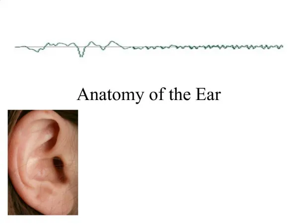
BASIC EAR ANATOMY. Major Divisions of the Ear. Divided into 4 parts (by function): External Ear Middle Ear Inner Ear Central Auditory Nervous System. Embryology of External Ear. SIX HILLOCKS OF HIS (WEEK 5)

E N D
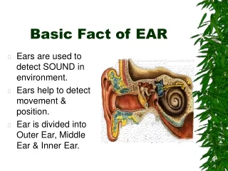
Major Divisions of the Ear Divided into 4 parts (by function): • External Ear • Middle Ear • Inner Ear • Central Auditory Nervous System
Embryology of External Ear • SIX HILLOCKS OF HIS (WEEK 5) • 6 TUBERCLES AROUND THE MARGIN OF FIRST VISCERAL CLEFT, DERIVED FROM 1st & 2nd BRANCHIAL ARCHES • TRAGUS; 1st ARCH • REST: REMAINING 5 TUBERCLES OF 2ND ARCH • FAULTY FUSION: PRE AURICULAR SINUS • ADULT SHAPE BY 20TH WEEK
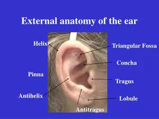
PINNA • EXTERNAL EAR • THE PINNA OR AURICLE • CONSISTS OF ELASTIC CARTILEGE AND SKIN • INCISURA TERMINALIS • ACTS AS A CHANNEL FOR SOUND WAVES AND DIRECTS THEM TOWARDS THE EXTERNAL AUDITORY MEATUS AND ON TO THE TYMPANIC MEMBRANE • Collects sound • Helps in sound localization • Most efficient in directing high frequency sounds to the eardrum • Source of graft material MADE UP OF 8 DISTINCT STRUCTURES
Auricle Nerve Supply Auriculotemporal Nerve (V) Lesser occipital Nerve Great auricular Nerve (C2 3, CNVII&X Great auricular Nerve CN VII &X 5
EXTERNAL AUDITORY CANAL: • DEVELOPMENT: • DEEPENING OF 1ST BRANCHIAL CLEFT, MEETS CORRESPONDING EVAGINATION FROM 1ST POUCH • RECANALISATION FROM 7TH MONTH FROM TM TOWARDS PHERIPHERY • FULLY DEVELOPED BY 28 WEEKS, COMPLETE BY 3 YEARS
External Auditory Canal • Bottom of concha - TM - 24mm. • Outer 1/3rd, cartilaginous - upwards, backwards & medially, • Inner 2/3rd, bony - downwards, forwards & medially. • To see TM - pull pinna upwards, backwards & laterally. 6
Cartilaginous part • •Outer 1/3rd-8mm-continous with cartilage of pinna • • Fissures of Santorini - parotid infections appear in meatus or vice cersa. • • Skin - thick, ceruminous & pilosebaceous glands -secrete wax. • CERUMEN (WAX) IS PRODUCED IN THE OUTER 1/3 AND IS A BACTERICIDAL WHICH AIDS IN THE CLEANING MECHANISM ALONG WITH EPITHELIAL MIGRATION • • Hair confined to outer canal - furuncles. 7
Bony part • inner 2/3rd - 16mm. — Skin - thin, continuous over TM, devoid of glands & hair follicles — 6mm lateral to TM - narrowing - Isthmus. — Ateroinferior part of deep meatus - Anterior Recess — Foramen of Huschke/tympanicum– Anteroinferior part deep canal, seen in children upto 5 years-infection to & fro from Parotid and TM joint. 8
Relations of External Auditory Canal • Superiorly - middle cranial fossa. • Posteriorly - mastoid air cells & FN • Inferiorly - parotid gland. • Anteriorly - temporomandibular joint. • Posterosuperior part of deep meatus -related to mastoid antrum - mastoiditis -sagging. 9
External Auditory Canal Nerve Supply - • Anterior wall & roof - auriculotemporal N - V3 • Posterior wall & floor - auricular branch of vagus - X - Arnolds N. • Posterior wall - sensory fibres of VII. 11
Tympanic Membrane Long crus of incus Posterior mallear fold Pars flaccida Anterior mallear fold Lateral process of malleus Pars tensa Manu briu m of malleus Umbo Cone of light %ia’' Right tympanic membrane (eardrum) viewed through speculum 12
Tympanic Membrane • Partition b/w EAC & ME. • Oblique - posterosuperior part is more lateral. Pearly white, cone of light anteroinferirorly • 9-10mm tall, 8-9mm wide & 0.1mm thick. • Pars Flaccida(Sharpnell's membrane) - above lateral process of malleus & b/w notch of rivinus and anterior & posterior malleal folds. . 13
Tympanic Membrane • Pars Tensa - most of TM. — Periphery thickened - fibrocartilaginousring -Annulus Tympanicus. — Central part tented inwards at tip of HOM -Umbo. — Cone of light - tip of HOM to periphery in AI Quad. 14
Tympanic Membrane • Three layers — Outer epithelial layer — Inner mucosal layer. — Middle fibrous layer - radial, circular & parabolic fibers. 15
Tympanic membrane BLOOD SUPPLY: OUTER/INNER SURFACE VEINS: OUTER/INNER SURFACE NERVE SUPPLY: OUTER – ANT: Auriculo temporal POST: ARNOLD`S INNER: TYMPANIC PLEXUS APPLIED ASPECTS: 1] HORIZONTAL IN NEW BORN 2] PARS FLACCIDA: RETRACTION POCKET 3] MYRINGOTOMY: 4] PERFORATIONS
MIDDLE EAR CLEFT • DEFINITION: • Aerated anteroinferiorly by ET, and posteriorly through aditus by the antrum of the mastoid and petrous temporal-bone
Middle Ear Cross section of ear including the middle ear 18
Embryology of Middle Ear: Tympanomastoid compartment • 3rd wk – tubotympanic recess (1st branchial pouch endoderm) approaches 1st cleft ectoderm • 6th wk – Ossicular mesenchyme separates cleft and pouch • 20th wk – tympanic cavity grows to enclose ossicles • 22nd wk – extension to form mastoid antrum • 33rd wk – early mastoid pneumatization
Middle Ear — Mesotympanum - level of PT — Epitympanum - Attic - above PT — Hypotympanum - below PT • Six sided box. 19
Middle Ear Structures: Boundaries of Tympanic Cleft • Roof: tegmen tympani—beyond is middle cranial fossa • Floor: jugular bulb • Anterior: ET orifice, carotid wall, tensor tympani canal, Canal of Huguier & Glasserian fissure • Posterior: Pyramidal eminence, facial nerve, aditus • Lateral: TM
Middle Ear • The Posterior Wall — Aditus - Attic communicates with Antrum. — bony projection - Pyramid - tendon of stapedius. — FN - runs behind pyramid. — Facial recess - FN, Chorda tympani, Fossa incudis. — Bony outer Attic Wall - Scutum. 21
Middle Ear • The Medial Wall - By Labyrinth — Bulge - Promontory - Basal coil of Cochlea — Oval Window - Foot plate of Stapes. — Round Window - Secondary TM. — Canal for facial N. — Prominance of lateral SCCanal. — Processuscochleariformis — Sinus tympani - deep recess medial to pyramid - Subiculum below & Ponticulus above. Fig. 1.7 Medial wall of middle ear. 1. Promontory 7. Ponticulus 2. Processuscochleariformis 8. Sinus tympani 3. CNVII 9. Subiculum 4. Oval window 10. Round window 5. Horizontal canal 11. Tympanic plexus 6. Pyramid 23
Contents of Middle Ear • •Chain of 3 small movable bones - Mallues, Incus and stapes • • 2 muscles – Tensor tympani and stapedius • 2 nerves - chorda tympani and the tympanic plexus of nerves. • Ligaments, Joints and mucosal folds • Air • 31
Ossicles • The Malleus — head, neck, handle, a lateral & an anterior process. — Head & neck in Attic. — Handle in fibrous layer of TM. — Lateral process - attachment to anterior & posterior malleal folds. The Incus - a body & a short process in attic, & a long process attaches to head of stapes. • The Stapes - head, neck, anterior & posterior crura & a footplate - oval window. 32
MUSCLES OF MIDDLE EAR - Dampen sound Auditory Muscles —Tensor Tympani - to neck of malleus -tenses TM - branch of mandibular N. - Stapedius - to neck of stapes - dampen loud sounds - Facial N
• Chorda tympani N - - Branch of FN enters ME through posterior canaliculus, runs on medial surface of TM b/w handle of malleus & long process of incus. - Taste from anterior 2/3rd of tongue, - secretomotor to submandibular & sublingual glands. 36
Tympanic plexus On promontory & formed by 1. Tympanic branch of Glossopharyngeal nerve 2. sympathetic fibers from plexus of internal carotid artery Tympanic plexus supplies • Lining membrane of tympanic cavity, mastoid air cells and auditory tube. • The lesser superficial petrosal nerve - otic ganglion -auriculotemporal nerve - secretomotor to Parotid. 37
Lining of ME Cleft • Mucous membrane of Nasopharynx is continuous with that of ME, Aditus, Antrum & air cells. • Wraps ME structures • Histologically - — ET - ciliated pseudostratified columnar in cartilaginous part & columnar in bony part, mucus glands in submucosa — Tympanic cavity - ciliated clomnar in anterior & inferior parts, cuboidal inposterior part. — Epitympanum & mastoid- flat nonciliated epithelium 38
Blood supply of ME • Anterior tympanic branch of maxillary artery. • Stylomastoid branch of posterior auricular artery. • Petrosal branch of middle meningeal artery. • Superior tympanic branch of middle meningeal artery. • Branch of artery of pterygoid canal. • Tympanic branch of internal carotid. 39
Lymphatic drainage • ME - retropharyngeal & parotid nodes. • ET - retropharyngeal nodes. 40
Facial nerve leaves Post. Cranial fossa via Internal Aud.Meatus - enters facial canal Intracranial, Intratemporal & extratemporal 1. Greater Petrosal N. GVE parasymp to Lacrimal gland, mucous glands of nose and palate, GVA sens. to Nasopharynx 2. Stapedial N. - SVE motor to stapedius 3. Chorda Tympani SVA taste to ant 2/3 tongue GVE parasymp to submandibular, subling. salivary glands
Eustachian Tube Mucous-lined, connects middle ear cavity to nasopharynx 36 mm long “Equalizes” air pressure in middle ear Normally closed, opens under certain conditions May allow a pathway for infection Children “grow out of” most middle ear problems as this tube lengthens and becomes more vertical
1*0*1. Root of /> ^ mt Supramaatoid Cre«x Parietal jTdtCh :- \ Zygoma .‘.laatoiJ Foamn ' \ v Ant. Root of Zygoma % V »«* «---- ill. > (Kminent. Artie.) P Of tl Of) A'( Part of r. rn.Vx] Fo*va Mattoid Pot Part of Glenoid F Laicrual Auditory Meats** (IMNN Plate) rNOM 26
Development of Mastoid • From Squamous & Petrous bones. • Petrosquamal suture may persist as a bony plate - Korner's Septum - Superficial squamous cells from deep petrous cells. • Difficulty in locating antrum & deeper cells. 30
Mastoid Process of Temporal Bone • Bony ridge behind the auricle, absent at birth • Develops from squamous & petrous • Korner`s septum • Hardest bone in body, protects cochlea and vestibular system • Provides support to the external ear and posterior wall of the middle ear cavity • Contains air cavities which can be reservoir for infection • MacEwen`s triangle • Air cell groups : 80% well pneumatized • Theories: Tumarkin,Diamant & Dahlberg, & Wittmack
Mastoid Antrum • Large air containing space in upper part of Mastoid, communicating with Attic through Aditus. • Roof - Tegmen antri - Middle cranial fossa. • Lateral wall - plate of bone 1.5cm thick -marked externally by suprameatal (Mac Ewen's) Triangle. 25
Aditus ad Antrum • Attic communicates with Antrum. • Prominance of Horizontal SCCon its medial side. • Fossa incudis - laterally. 27
Mastoid & its Air Cell System • Mastoid - bone cortex with a "honeycomb" of air cells underneath. • Depending on air cell development - - Well pneumatised / cellular - Diploetic - marrow spaces & few air cells. - Sclerotic / acellular 28
Mastoid Air Cell System • Depending on location - — Zygomatic cells. -squamous cells — Tegmen cells. — Perisinus cells. — Retrofacial cells. — Perilabyrinthine cells. — Peritubal cells. — Tip cells 29