Download

1 / 17
330 likes | 1.3k Views
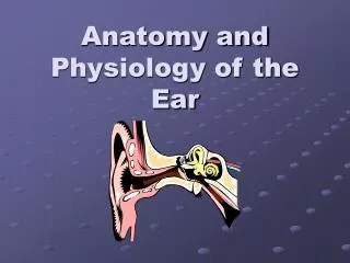
Anatomy of the Ear. Miss Martini’s 7 th Grade Science Class. Click to start. Click on the each part of the ear To learn more . QUESTION. The Cochlea. The spiral cavity of the inner ear containing the organ of Corti The cochlea produces nerve impulses in response to sound vibrations.

E N D
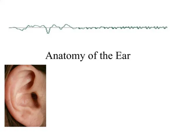
Anatomy of the Ear Miss Martini’s 7th Grade Science Class Click to start
The Cochlea • The spiral cavity of the inner ear containing the organ of Corti • The cochlea produces nerve impulses in response to sound vibrations Go back
The Cochlear Nerve • The branch of the auditory nerve that connects with the cochlea and transmits impulses to the hearing center of the brain Go back
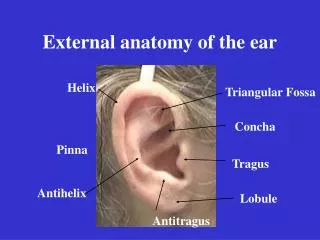
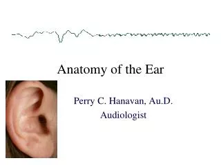
The Auricle • The projecting, outer part of the ear • The auricle is also known as the pinna Go back
The Tympanic Membrane • A membrane forming part of the organ of hearing, which vibrates in response to sound waves • The tympanic membrane forms the eardrum, between the outer and middle ear Go back
The External Meatus • The external meatus is a tube running from the outer ear to the middle ear. The human ear canal extends from the pinna to the eardrum and is about 26 mm in length and 7 mm in diameter • The external meatus is also known as the ear drum Go Back
The Incus • One of the three tiny bones in the middle ear • Sound impulses coming from the eardrum are conveyed from the malleus to the incus and from the incus to the stapes at the oval window to the inner ear Go Back
The Malleus • The hammer-shaped bone that is the outermost of the three small bones in the mammalian middle ear • The malleus can also be called the hammer Go back
The Stapes • The innermost of the three small bones of the middle ear • The stapes can also be called the stirrup Go back
The Auditory Tube • The tube that runs from the middle ear to the pharynx • The function of this tube is to protect and drain the middle ear • Can also be known as the Eustachian tube Go back
The Semicircular Canals • The three looped fluid-filled membranous tubes, at right angles to one another • The semicircular canals control the senses of orientation and equilibrium Go back
QUESTION • Which is located between the outer and middle ear, forming the eardrum? • A. The Cochlea • B. The Malleus • C. The Tympanic Membrane
Uh-Oh! • Sorry, not the Cochlea! • The Cochlea is located in the inner ear and responds to sound vibrations! TRY AGAIN!
Uh-Oh! • Sorry, not the Malleus! • The Malleus is the hammer shaped bone in the middle ear! Try again!
Good Job!!! • Correct! • The Tympanic Membrane is located between the outer ear and the middle ear, forming the ear drum! Continue
The End! • You have successfully completed the Anatomy of the Ear tutorial! • GREAT JOB!!! • Please click “back to the beginning” for the next student! Back to the beginning Good Job!