Download
1 / 28
370 likes | 817 Views
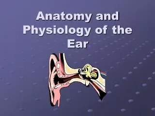
Anatomy of the Ear. Parts of the Ear. Outer Ear: From the pinna to the tympanic membrane . Middle Ear: From the tympanic membrane to the end of the stapes . Inner Ear: From the oval window to the auditory nerve. The Outer Ear.

E N D
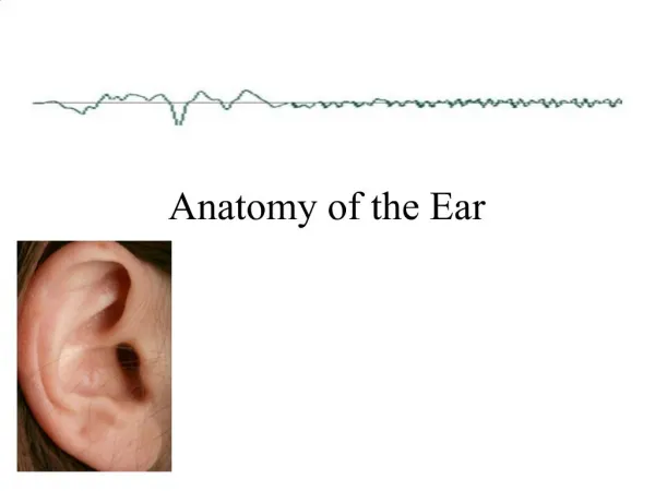
Parts of the Ear Outer Ear: From the pinna to the tympanic membrane. Middle Ear: From the tympanic membrane to the end of the stapes. Inner Ear: From the oval window to the auditory nerve.
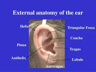
The Outer Ear • The first part of the outer ear is the pinna or the auricle. • The purpose of the pinna is to collect and locate sound. Pinna Crus of Helix Concha Lobule
The Outer Ear • The external auditory meatus is also known as the Ear Canal. • The purpose of the external auditory meatus is to channel sound to the middle ear. • Cerumen or wax is produced in this part of the ear. External Auditory Meatus
Tympanic Membrane • The tympanic membrane is the dividing point between the middle and outer ear. • The tympanic membrane (TM) vibrates to carry the sound to the middle ear. • It is made of tissue similar to skin. • The tympanic membrane is also called the ear drum.
The Middle Ear • The middle ear is an air filled cavity. • The ossicular chain is the name for three bones in the middle ear used for hearing. • The Eustachian tube is also in the middle ear.
The Ossicular Chain • The ossicular chain, also called theossicles, is a series of three bones. • Malleus, Incus, Stapes • These bones are the smallest bones in the body. • The bones are all connected. • The TM vibrates the malleus and the sound energy travels through the incus and the stapes. The stapes is connected to the oval window.
The Ossicular Chain Here are the bones in the ossicular chain. In the ear they are connected.
The Eustachian Tube • A tube that connects the middle ear to the throat. • The purpose is to regulate air pressure in the ear. • It allows fluid from behind the ear drum to be discharged into the back of the nose.
The Inner Ear • The inner ear is comprised of the cochlea, and the semicircular canals. • The stapes is connected to the oval window. • The oval window is the opening to the cochlea. • The cochlea is the organ used for hearing. • The semicircular canals are used for balance.
The Cochlea • The cochlea is a snail-shaped organ filled with tiny hairs used for hearing. • It is connected to the auditory nerve.
Inside the Cochlea • The cochlea is a long tube curled up like a snail shell. • This tube is divided into three sections. • Each section is filled with fluid. • In the middle section, is the Organ of Corti. • This organ is the organ that we hear with.
Organ of Corti • This energy is transferred to the fluid. • This fluid stimulates hairs on the Organ of Corti. • This sends an impulse to the auditory nerve.
The Semicircular Canals • The semicircular canals control balance. Semicircular Canals Oval Window Cochlea Round Window
Review This picture demonstrates the chain of events that happens when a sound is heard. Do not worry about the specific names.
Otitis Media • Otitis Media, also known as an ear infection, is a common infection of the middle ear cavity. • This happens when the Eustachian tube becomes blocked and the middle ear cavity becomes filled with fluid.
Otitis Media • A doctor can tell if a person has otitis media by using an otoscope to look down a person’s ear canal and looking at the tympanic membrane. • If the doctor sees the “cone of light” then the person’s ear is not infected. • If the doctor does not see the light and the tympanic membrane is cloudy, the person probably has otitis media. Healthy Tympanic Membrane Otoscope Otitis Media Cone of light
Other Problems with the Ear • Otolaryngologist • Also known as an ear, nose, and throat (ENT) specialist • This doctor can diagnose problems that can occur in the ear • Some other problems besides otitis media are: • Presence of foreign bodies • Impacted cerumen (wax) • External otitis (swimmer’s ear) • Perforation of TM (tear in the TM) • Otalgia (earache)
Describing a Hearing Loss • There are three ways to categorize hearing loss. • Type • Degree • Age at Onset
Type of Hearing Loss • There are three types of hearing loss. • Sensorineural • Conductive • Mixed • All three have an impact on a person’s hearing. • See page 7 in the course pack.
Conductive Loss • A person has a conductive loss if they have any problem with the outer or middle ear. • For example, if a person is born without a pinna or if a person has severeotitis media. • A conductive loss is almost always “fixable.” • Meaning that if you have otitis media and you take antibiotics then your conductive loss will go away, or if you born without a pinna, you can have one constructed. The part of the picture shaded in purple, shows where a conductive loss is located.
Sensorineural Loss • A sensorineural loss is a hearing loss that involves problems with the cochlea and/or the auditory nerve. • For example, sometimes the hairs in the Organ of Corti are damaged, destroyed, or missing. This causes a sensorineural hearing loss. • The abbreviation often used for a sensorineural hearing loss is SNHL. • A sensorineural hearing loss cannot be “fixed.” It can only be helped by hearing aids and cochlear implants.
Mixed Hearing Loss • A mixed hearing loss is where a person has both a conductive and a sensorineural loss. • For example, when a person has otitis media and has damaged hair cells. This would cause both a conductive and a sensorineural hearing loss. • When the person “fixes” the conductive hearing loss, then the person just has a sensorineural hearing loss.
Degree of Hearing Loss • The degree of hearing loss is determined by what a person can or cannot hear. • To determine this, a person goes to an audiologist and has a hearing test done. • The result of the test is shown on an audiogram. • An audiogram is a visual representation of a person’s hearing. • An audiogram shows a person’s “thresholds” or the softest sound a person can hear at various frequencies.
Audiograms • On the left side of the audiogram is decibels (dB) or the loudness of the sound. • On the top is the frequency which is measured in Hertz (Hz). The frequency measures the pitch of the sound. • The markings for the right ear are a red circle, O To remember this, think red-right-round. • The markings for the left ear are blue X’s. • Please see page 9 in your course pack.
Degree of Hearing Loss • Normal Hearing: 25 dB and below • Mild Hearing Loss: 25-40 dB • Moderate Hearing Loss: 40-55 dB • Moderately Severe Loss: 55-70 dB • Severe Loss: 70-90 dB • Profound Loss: 90+
Degree of Hearing Loss • Normal Hearing: 25 dB and below • Mild Hearing Loss: 25-40 dB • Moderate Hearing Loss: 45-55 dB • Moderately Severe Loss: 55-70 dB • Severe Loss: 70-90 dB • Profound Loss: 90+ The specific numbers may vary. Different sources and professions use different numbers. These are just one set of numbers.
Age at Onset • When a person acquires a hearing loss is called the age at onset. • There are two periods when this can occur: • Pre-lingual: Before language is developed • Post-lingual: After language has developed • Why do you think it is important as teachers to know when your students acquired their hearing loss?