Download
1 / 39
650 likes | 2.04k Views
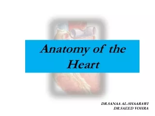
Heart Anatomy . Size, Location, and Orientation Enclosed in the mediastinum Base (posteriorsuperior portion) and Apex (inferioranterior portion) . Heart Anatomy. Coverings Pericardium protects the heart anchors the heart to surrounding structures such as the diaphragm and the great vessels prevents overfilling of the heart with blood .

E N D
1.
Anatomy & physiology
of the heart
4. Heart Anatomy
Size, Location, and Orientation
Enclosed in the mediastinum
Base (posteriorsuperior portion) and Apex (inferioranterior portion)
5. Heart Anatomy Coverings
Pericardium
protects the heart
anchors the heart to surrounding structures such as the diaphragm and the great vessels
prevents overfilling of the heart with blood
6. Heart Anatomy Coverings
pericardial cavity contains a film of serous fluid
pericarditis: inflammation of the pericardium which may lead to adhesions between the layers or the buildup of fluid in the pericardial cavity (cardiac tamponade)
7. Heart Anatomy Heart Wall
Epicardium
Myocardium
bulk of the heart consisting mainly of cardiac muscle
8. Heart Anatomy Heart Wall
Endocardium
simple squamous epithelium and a thin CT layer that lines the heart chambers and valves and is continuous with the endothelial lining of the blood vessels
9. Heart Anatomy Chambers
Atria
Features
small, thin-walled chambers
Functions
receiving chambers for blood returning to the heart from the circulation
push the blood into the adjacent ventricles.
10. Heart Anatomy Chambers
Atria
Receive blood from
right side
Superior and Inferior Vena Cava
Coronary Sinus (draining the myocardium)
left side
Pulmonary Veins
11. Heart Anatomy Chambers
Ventricles
Features
make up most of the mass of the heart
the walls of the left ventricle are 3X thicker than those of the right
12. Heart Anatomy Chambers
Ventricles
Functions
discharging chambers of the heart
propel blood to Pulmonary Trunk (right ventricle), Aorta (left ventricle)
14. Heart Anatomy Pathway of Blood Through the Heart
Pulmonary Circuit
functions strictly as gas exchange
the right side of the heart is the pulmonary circuit pump
this is a short, low-pressure circuit
15. Heart Anatomy Pathway of Blood Through the Heart
Systemic Circuit
functions as both gas and nutrient exchange
the left side of the heart is the systemic circuit pump
this is a long, high-resistance pathway through the entire body
16. Heart Anatomy Heart Valves
These enforce the one-way flow of blood through the heart
The valves open and close in response to differences in blood pressure on their two sides
17. Heart Anatomy Heart Valves
Atrioventricular Valves
the valves close when the ventricular pressure increases and forces blood against the valve flaps
Tricuspid (right side)
Bicuspid (Mitral) (left side)
18. Heart Anatomy Heart Valves
Semilunar Valves
located between the ventricles and the large arteries
these open when the pressure produced by the contracting ventricle exceeds that in the artery and close when the arterial pressure exceeds the pressure produced by the relaxing ventricle
Pulmonary (right side)
Aortic (left side)
20. Coronary Circulation Coronary Arteries
the coronary arteries arise from the base of the aorta and actively deliver blood only when the heart is relaxed
the heart is 0.5% of body weight and receives 5% of the body's blood supply (most to the left ventricle)
21. Coronary Circulation Coronary Arteries
left main coronary artery
left anterior descending artery: serves the interventricular septum and anterior walls of both ventricles
circumflex artery: serves the left atrium and posterior wall of the left ventricle
22. Coronary Circulation Coronary Arteries
Right main coronary artery
posterior descending artery: serves the posterior walls of both ventricles
marginal artery: lateral wall of the right side of the heart
Cardiac Veins follow arteries and join at the Coronary Sinus which empties blood into the right atrium
24. Heart Physiology Electrical Events
Intrinsic Conduction System of the Heart
the ability of cardiac muscle to depolarize and contract is intrinsic (no nervous stimulation is required)
nerve impulses can alter the basic rhythm of heart activity set by intrinsic factors
25. Heart Physiology Electrical Events
Action Potential Generated by Autorhythmic Cells
Sequence of Excitation
Sinoatrial Node
Atrioventricular Node
Atrioventricular Bundle (bundle of His)
Bundle Branches
Purkinje Fibers
26. Heart Physiology Electrical Events
Extrinsic Innervation of the Heart
fibers of autonomic nervous system accelerate or inhibit the basic rate of heartbeat set by the intrinsic conduction system
27. Heart Physiology Electrical Events
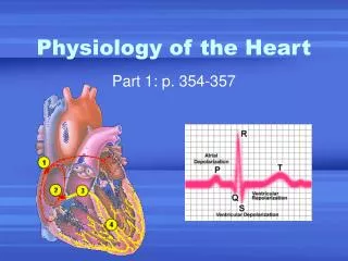
Electrocardiography
electrical currents generated and transmitted through the heart spread throughout the body and can be monitored
the graphic recording of electrical changes during heart activity is called an electrocardiogram (ECG or EKG)
28. Heart Physiology Electrical Events
Electrocardiography
the ECG consists of series of three waves
P Wave: atrial depolarization starting at the SA node
QRS Complex: ventricular depolarization
29. Heart Physiology Electrical Events
Electrocardiography
P-R (P-Q) interval: time from the beginning of atrial excitation to the beginning of ventricular excitation and includes the contraction of the atria and the passage of the depolarization wave through the rest of the conduction system
30. Heart Physiology Electrical Events
Electrocardiography
T Wave: ventricular repolarization
Q-T interval: time from the beginning of the ventricular depolarization through their repolarization and includes the contraction of the ventricles
31. Heart Physiology Mechanical Events: The Cardiac Cycle
Terms
Systole: contraction period of heart activity
Diastole: relaxation period of heart activity
32. Heart Physiology Mechanical Events: The Cardiac Cycle
Cardiac Cycle
pressure in the heart is low and the blood is returning passively (70% of ventricle filling occurs)
atria depolarize (P wave) and contract and force the remaining 30% of the blood into the ventricles
the atria relax and remain in diastole through the rest of the cycle
33. Heart Physiology Mechanical Events: The Cardiac Cycle
the ventricles depolarize (QRS complex)
ventricles begin their contraction
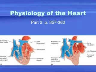
ventricular pressure rises rapidly and the AV valves close
as ventricular pressure rises above arterial pressure the semilunar valves open and the ventricles empty during the ventricular ejection phase
34. Heart Physiology Mechanical Events: The Cardiac Cycle
ventricular systole ends with the repolarization of the ventricles (T wave)
ventricles relax and ventricular pressure drops
semilunar valves close
the atria have been filling with blood since ventricular systole and when the atrial pressure exceeds the ventricular pressure the AV valves open ventricular filling begins again
35. Heart Physiology Cardiac Output
General
cardiac output is the amount of blood pumped out by each ventricle in 1 minute and is the product of heart rate (HR) and stroke volume (SV)
stroke volume is the volume of blood pumped out by one ventricle with each beat and is the difference between end diastolic volume (EDV) and the end systolic volume (ESV)
36. Heart Physiology Cardiac Output
Regulation of Stroke Volume
Preload: Degree of Stretch
affected by the EDV and operates intrinsically
Frank Starling Law of the Heart: The greater the degree of stretch of cardiac muscle fibers the greater the force of contraction and the greater the stoke volume
37. Heart Physiology Cardiac Output
resting cardiac fibers are normally shorter than the optimal length and stretching them (increasing EDV) produces dramatic increases in contractile force
anything that increases the volume or speed of venous return (slow heart rate or exercise) increases EDV which increases the force of contraction which increases stroke volume
38. Heart Physiology Cardiac Output
Contractility
affects the ESV and are extrinsic factors that increase the contractile strength of heart muscle
many chemicals enhance contractility (positive inotropic agents)
39. Heart Physiology Cardiac Output
Afterload: Back Pressure
affects the ESV
the pressure exerted on the aortic (80 mm Hg) and pulmonary (20 mm Hg) valves by arterial blood
important in people with hypertension where ESV is increased and stroke volume is reduced