Download
1 / 28
400 likes | 745 Views
Anatomy of the Heart. OBJECTIVES. At the end of the lecture, the student should be able to : Describe the shape of heart regarding : apex, base, sternocostal and diaphragmatic surfaces. Describe the interior of heart chambers : right atrium, right ventricle, left atrium and left ventricle.

E N D
OBJECTIVES • At the end of the lecture, the student should be able to : • Describe the shape of heart regarding :apex, base, sternocostal and diaphragmatic surfaces. • Describe the interior of heart chambers : right atrium, right ventricle, left atrium and left ventricle. • List the orifices of the heart : • Right atrioventricular (Tricuspid) orifice. • Pulmonary orifice. • Left atrioventricular (Mitral) orifice. • Aortic orifice. • Describe the innervation of the heart • Breifly describe the conduction system of the heart.
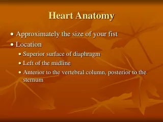
It lies in the middle mediastinum. It is surrounded by a fibroserous sac called pericardium which is differentiated into an outer fibrous layer (Fibrouspericardium) & inner serous sac (Serous pericardium). The Heart is somewhat pyramidal in shape, having: Apex Sterno-costal (anterior surface) Base (posterior surface). Diaphragmatic (inferior surface) It consists of 4 chambers, 2 atria (right& left) & 2 ventricles (right& left) The Heart
Apex of the heart Note that the base of the heart is called the base because the heart is pyramid shaped; the base lies opposite the apex. The heart does not rest on its base; it rests on its diaphragmatic (inferior) surface Directed downwards, forwards and to the left. It is formed by the left ventricle. lies at the level of left 5th intercostal space 3.5 inch from midline.
Sterno-costal (anterior)surface Divided by coronary (atrio-ventricular) groove into : Atrial part, formed mainly by right atrium. Ventricular part , the right 2/3 is formed by right ventricle, while the left l1/3 is formed by left ventricle. The 2 ventricles are separated by anterior interventricular groove, which lodges : Anterior interventricular artery (branch of left coronary). Great cardiac vein. The coronary groove lodges the right coronary artery. This surface is formed mainly by the right atriumand the right ventricle.
Sterno-costal (anterior)surface The marginal branch of right coronary artery runs along the inferior border The funnel-shaped part of right ventricle just below pulmonary trunk is called infundibulum Infundibulum
Diaphragmatic (Inferior)surface • Formed by the 2-ventricles, mainlyleftventricle (left 2/3). • Slightly concave as it rests on diaphragm. • Directed inferiorly & backward. • Separated from base of heart by posterior part ofcoronary sulcus • The 2-ventricles are separated by posterior interventricular groove which lodges: • Posterior interventricular artery • Middle cardiac vein
Base of the Heart (posterior surface) • It is formed by the 2 atria, mainlyleft atrium,into which open the 4 pulmonary veins. • It is directed backwards. • Lies opposite middle thoracic vertebrae (5-7). • Is separated from the vertebral column by descending aorta, esophagus and oblique sinus ofpericardium • Bounded inferiorly by post part of coronary sulcus , which lodges the coronary sinus
Borders of the Heart Upper border: Is formed by the 2 atria. It is concealed by ascending aorta & pulmonary trunk. Right border: Is formed by right atrium Lower border: Is formed mainly by right ventricle + apical part of left ventricle. Left border: Is formed mainly by left ventricle + auricle of left atrium.
Chambers of the Heart The heart is divided by vertical septa into four chambers: the right and left atria and the right and left ventricles. The right atrium lies anterior to the left atrium, and the right ventricle lies anterior to the left ventricle.
Right Atrium • The right atrium consists of a main cavity and a small out pouching, the auricle. • On the outside of heart at the junction between the right atrium and the right auricle is a vertical groove extending from the opening of SVC to the opening of IVC. • This is called the sulcus terminalis, which on the inside forms a ridge, the crista terminalis.
Cavity of Right Atrium • Crista terminalis divides right atrium into: 1- Anterior part:rough and trabeculated by bundles of muscle fibres (musculi pectinati). • 2- Posterior part (sinus venarum) : is smooth. • The interatrial septum carries an oval depression called Fossa ovalis. The margin of this depression is called Anulus ovalis. • The blood leaves right atrium to right ventricle via tricuspid valve.
Cavity of Right Atrium Openings in right atrium: • SVC --- has no valve • IVC --- guarded by a valve • Coronary sinus : has a well-defined valve • Right atrioventricular orifice lies anterior to IVC opening , it is surrounded by a fibrousring which gives attachment to the tricuspid valve • Small orifices of small veins
Right ventricle • The right ventricle communicates with the right atrium through the atrioventricular orifice. • It also communicates with the pulmonary trunk through the pulmonary orifice. • As the cavity approaches the pulmonary orifice it becomes funnel shaped, at which point it is referred to as the infundibulum. Infundibulum
Cavity of right ventricle • Its wall is thinner than that of left ventricle. • Its wall contains projections called trabeculae carnae. • The right ventricle communicates with right atrium through right atrioventricular orifice & with pulmonary trunk through pulmonary orifice. • Large projections arise from the walls called papillary muscles • Anterior papillary muscle • Posterior papillary muscle • Septal papillary muscle
Cavity of right ventricle • Each papillary muscle is attached to the cusps of tricuspid valve by tendinous threads called chordae tendinae. • Blood leaves the right ventricle to pulmonary trunk through pulmonary orifice. • The wall of infundibulum is smooth and contains no trabeculae. • Interventricular septum is connected to anterior papillary muscle by a muscular band called moderator band
Right atrio-ventricular (tricuspid) orifice • About one inch wide, admitting tips of 3 fingers. • It is guarded by a fibrous ring which gives attachment to the cusps of tricuspid valve. • It has 3-cusps (anterior-posterior-septal or medial). • The atrial surface of the cusps are smooth, while their ventricular surfaces give attachment to the chordae tendinae.
Pulmonary orifice • Surrounded by a fibrous ring which gives attachment to the cusps of the pulmonary valve. • The valve is formed of 3 semilunar cusps :2 anterior and one posterior which are concave superiorly and convex inferiorly. • Nochordae tendineae or papillary muscles are attached to these cusps
Left atrium of the heart • It communicates with the left ventricle through the atrioventricular orifice • It forms the greater part of base ofheart. • Its wall is smooth except for small musculi pectinati in the left auricle. • Recieves 4 pulmonary veins which have no valves. • Sends blood to left ventricle through the left atrioventricular orifice which is guarded by mitral valve.
Left ventricle of the heart • Its wall is thicker than that of right ventricle. • It receives blood from left atrium through left atrio-ventricular orifice which is guarded by mitral valve. • Its wall contains trabeculae canae. • Its wall contains 2 large papillary muscles (anterior & posterior). They are attached by chordae tendinae to cusps of mitral valve.
Left ventricle of the heart • The blood leaves the left ventricle to the ascending aorta through the aortic orifice. • The part of left ventricle leading to ascending aorta is called aortic vestibule. The wall of this part is fibrous and smooth.
Left atrio-ventricular (mitral) orifice • Smaller than the right, admitting only tips of 2 fingers. • Guarded by a mitral valve. • Surrounded by a fibrous ring which gives attachment to the cusps of mitral valve. • Mitral valve is composed of 2 cusps: • Anterior cusp : lies anteriorly and to right. • Posterior cusp : lies posteriorly and to left. • The atrial surfaces of the cusps are smooth, while ventricular surfaces give attachment to chordae tendinae.
Aortic orifice • Surrounded by a fibrous ring which gives attachment to the cusps of aortic valve. • Aortic valve is formed of 3 semilunar cusps which are similar to those of pulmonary valve, but the position of the cusps differs being one anteriorand 2 posterior.
Nerve supply of the heart By sympathetic & parasympathetic fibers via the cardiac plexus situated below arch of aorta. The sympathetic fibres arise from the cervical & upper thoracic ganglia of sympathetic trunks. The parasympathetic fibres arise from the vagus nerves. Postganglionic fibres reach heart along – SAN ,AVN & nerve plexus around coronary arteries. Sympathetic Fs.--- accelerate heart rate but Parasympathetic Fs --- slow heart rate (constriction of coronay arteries)
Conduction system of the heart • The beating of the heart is regulated by the intrinsic conduction (nodal) system • Its function is to ensure that the chambers of the heart contract in the proper rhythm and sequence. • The main center is the sinoatrial (SA) node, located in the right atrium • The atrioventricular (AV) node is located at the junction of the atria and the ventricles
Conduction system of the heart The atrioventricular (AV) bundle (bundle of His) is located in the interventricular septum The Purkinje fibers are located inside the walls of the ventricles the SA node is called the pacemaker of the heart, because it generates the impulse
Conduction system of the heart The heart contractionbegins with an electrical impulse in the SA node. The impulse spreads to the two atria and triggers their contraction. Then it reaches the AV node After that, the impulse travels along the AV bundle At the end, it reaches the Purkinje fibers in the walls of the ventricles and triggers their contraction Thus, the conduction system of the heart ensures simultaneous contraction of the atria and the ventricles