Download
1 / 23
430 likes | 911 Views
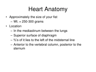
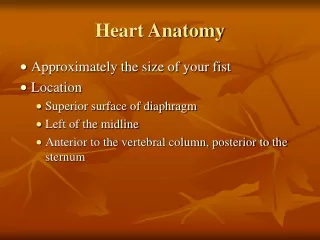
ANATOMY OF HEART. Thorax Thorax is the superior part of the trunk between the neck and abdomen. It extends below the neck to the diaphragm. It contains the primary organs of respiratory and cardiovascular system.

E N D
Thorax • Thorax is the superior part of the trunk between the neck and abdomen. • It extends below the neck to the diaphragm. • It contains the primary organs of respiratory and cardiovascular system
A typical human rib cage consists of 24 ribs, the sternum, costal cartilages, and the 12 thoracic vertebrae. • All ribs are attached in the back to the thoracic vertebrae.
The upper seven are true ribs, are attached in the front to the sternum by means of costal cartilage. Due to their elasticity they allow movement when inhaling and exhaling. • The 8th, 9th, and 10th ribs are called false ribs, and join with the costal cartilages of the ribs above. • The 11th and 12th ribs are known as floating ribs, as they do not have any anterior connection to the sternum. • The spaces between the ribs are known as intercostal spaces; they contain the intercostal muscles, nerves, and arteries.
The thoracic cavity is divided into three major spaces. • The central/median compartment called mediastinum houses the conducting structures(esophagus,trachea,major blood vessels and most importantly heart).
Pericardium There are two layers to the pericardial sac: • the fibrous pericardium and • the serous pericardium. • The serous pericardium, in turn, is divided into two layers, the parietal pericardium and visceral pericardium
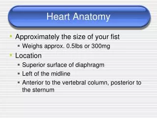
Heart The wall of each heart chamber consists of • Endocardium:thin internal layer of endothelium • Myocardium: thick, middle layer composed of cardiac muscle • Epicardium:thin external layer formed by the visceral layer of serous pericardium
The heart has four chambers: • Right and left atria • Right and left Ventricles
Atrium Right atrium • The superior vena cava opens at the level of R. 3rd costal cartilage and inferior vena cava at 5th costal cartilage. • The right atrium opens into the R. ventricle thru Tricuspid valve • Receives poorly oxygeneted blood from superior vena cava, inferior vena cava and coronary sinus
Left atrium • The pairs of right and left pulmonary veins enter the posterior aspect of atrium. • The left atrium opens into L. ventricle thru Bicuspid or mitral valve. • Receives rich oxygenated blood from pulmonary veins from the lungs
Ventricles Right ventricle: • Receives the deoxygenated blood from the atrium • And thru R. and L. pulmonary arteries pumps blood to the lungs Left ventricle: • Receives the oxygenated blood from the atrium • And thru Aorta pumps blood to the systemic circulation • Left ventricle more thicker than the right ventricle
Papillary muscles • Papillary muscles are nothing but the conical muscular projections from the myocardium of the heart • They support, strenghthen and responsible for the opening and closure of the cuspid valves. • They area attached to the cuspid valves through tendinous cords called chordaetendinae
Conducting system of the heart • Conducting system of the heart consists of the cardiac muscle cells, SA and AV nodes, purkinjefibres and Bundle of His.
Sinuatrial node(SA) • It is located antero-laterally just deep to epicardium at the jucnction of Superior Venacava and R.atrium. • It is the pacemaker of the heart, initiating and regulating the impulses for contraction. • It is a small collection of nodal tissue and specialized cardiac muscle fibres. • It could be stimulated or inhibited by the sympathetic or parasympathetic division
Atrio-Ventricular Node(AV) • Smaller collection of nodal tissue than SA node. • Located in the posteroinferior region of the interatrial septum.
Bundle of His • AV bundle- the AV node passes the signal from atrium to the ventricles thru AV bundle, which is the only bridge between the atrial and ventricular myocardium. • Av bundle divides into R. and L. bundle • These bundles proceed on each side and then ramify into purkinjefibres • These purkinjefibres on both side stimulate the papillary muscles and respective ventricles.