Download
1 / 32
330 likes | 398 Views
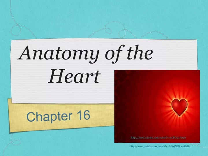
Anatomy of the Heart. Chapter 16. http://www.youtube.com/watch?v=nCPOio1FQ5Q. http://www.youtube.com/watch?v=AOiyjNFB0as&NR=1. Cardiology = study of the heart.

E N D
Anatomy of the Heart Chapter 16 http://www.youtube.com/watch?v=nCPOio1FQ5Q http://www.youtube.com/watch?v=AOiyjNFB0as&NR=1
Cardiology = study of the heart • Function: to pump and force blood through the blood vessels of the body, providing every cell in the body with vital nutrients and oxygen • Heart: hollow muscular organ • Sits in the chest within the mediastinum, between the lungs • Apex - lower,pointed end of the heart, located at the level of the fifth intercostal space • Base - upper, flat portion of the heart, located at the level of the second rib • Precordium - area of the anterior chest wall overlying the heart and the great vessels • Anatomical location - used for evaluating heart sounds, applying electrodes for EKG, and cardiopulmonary resuscitation (CPR)
Layers and Coverings of the heart • Layers: • Endocardium - inner most layer, lines the valves and is continuous with the blood vessels that enter and leave the heart - smooth shiny surface allows blood to flow easily • Myocardium - middle layer, thick, composed of cardiac muscle that contracts an pumps blood through the blood vessels • Epicardium - thin outer most layer of the heart, helps to form the pericardium (visceral pericardium)
Pericardium • Pericardium - support the heart, sling-like structure, attaches the heart to the surrounding structures (diaphragm, great vessels) layers: • epicardium (visceral pericardium) - lines the heart and folds back to form the parietal pericardium • pericardial space - pericardial membranes are serous - secrete small amount of slippery serous fluid (10-30 ml) - lubricates and decreases friction • parietal pericardium - attaches to the outer fibrous pericardium • Fibrous pericardium - anchors the heart to surrounding structures • Pericarditis - inflammation of pericardial membranes - characterized by pain, friction rub (heart sound) • Pericardial effusion - associated with pericarditis - when the inflamed membranes secrete excess serous fluid into the pericardial space - compresses the heart making it difficult to for the heart to relax and fill with blood • Cardiac tamponade - when there is an accumulation of either fluid/blood in the pericardial space compressing the heart to a point of insufficient pumping of blood to the body - life-threatening condition • treatment- insertion of a long needle into the pericardial space to aspirate (suck-out) excess fluid/blood
Double Pump - beats as one!Two circulations • Right heart- rt side of the heart • receives unoxygenated blood from the superior and inferior vena cava (large veins that collect blood from the body) • right heart pumps blood to he lungs where it gets oxygenated • Pulmonary circulation- the path that the blood follows from the right side of the heart to the lungs and back to the left side of the heart • function: is to pump blood through the lungs in order to pick up oxygen and get rid of carbon dioxide • Left heart - left side of the heart • received oxygenated blood from the lungs and pumps it to all the organs of the body • Systemic circulation- the path that the blood follows from the left heart to all the organs and back to the right side of the heart • Septa- wall that separates the chambers • Interatrial septum - separates right and left atria • Interventricular septum - separates right and left ventricles
The Hearts Chambers • Four Chamber: 2 atria, 2 ventricles • Atrium - upper chambers - receive blood into the heart • Ventricle - lower chambers and pump blood out of the heart, thick myocardial layer, thick muscle is needed to create enough force to pump blood out of the heart • Right Atrium - thin walled cavity, receives unoxygenated blood from the superior and inferior vena cava • Superior vena cava - collects unoxygenated blood from the head and upper body region • Inferior vena cava - collects unoxygenated blood from the lower part of the body • Right Ventricle - receives unoxygenated blood from the right atrium, pumps blood through the pulmonary arteries to the lungs • Left Atrium - thin walled cavity that receives oxygenated blood from the lungs through 4 pulmonary veins • Left Ventricle - receives oxygenated blood from the left atrium, pumps blood into the systemic circulation through the aorta (largest vessels in the body) • myocardial layer if the left ventricle is thicker than the right - left ventricle has to pump against a greater amount of force in order to efficiently pump blood into the systemic circulation -
Ventricular Hypertrophy • Ventricular Hypertrophy - when the ventricle is forced to overwork it becomes enlarged • high BP (chronically hypertensive) - the pressure in the aorta makes it more difficult for the left ventricle to pump blood into the aorta - the ventricle works harder and enlarges - • if untreated - the ventricle will eventually fail as a pump • Right Ventricular hypertrophy - for the same reason the right ventricle will become enlarged if the pulmonary artery pressure is too great and lead to right heart failure.
Great Vessels of the Heart • Great Vessels - are large blood vessels attached to the heart... • Superior vena cava • Inferior vena cava • Pulmonary artery • Pulmonary veins (4) • Aorta
Heart Valves • 4 valves • function: is to keep blood flowing in a forward direction, are in the entrance and exit of the chambers • AV valves (atrioventricular valves)- located between atria and ventricles • right AV valve (tricuspid valve) • left AV valve (bicuspid valve) • Semilunar valves - control outflow of blood from the right and left ventricles • pulmonic semilunar valve (right semilunar valve) • aortic semilunar valves (left semilunar valve)
AV Valvesatrioventricular valves • Location: between the atria and ventricles • AV valves have cusps/flaps that hang loose when relaxed - valves are open and allows blood flow from the atria to the ventricle • what closes the AV valve? .....pressure!! • when the ventricles contract, the heart muscle compresses and squeezes the blood...blood is forced behind the cusps/flaps of the AV valves and pushes them toward the atria - closing the valve ...preventing the back-flow of blood into the atria • Chordaetendineae - tough fibrous bands of tissues attached to the AV valve and the ventricular wall - prevents the AV valve from being pushed up into the atria when the ventricle contracts and closes the AV valve • Right AV valve (tricuspid) - located between the right atrium and ventricle • has three cusps = tricuspid , that allow blood flow from the right atrium to right ventricle when the valve is open and prevent back-flow of blood from the right ventricle to the right atrium when the valve is closed • Left AV valve (bicuspid) (mitral valve) - located between left atrium and left ventricle • has two cusps = bicuspid, mitral valve - when open allow blood flow from the left atrium into the left ventricle, when Left AV valve is closed prevents blood flow from the left ventricle into the left atrium
Semilunar Valvessemi=half, lunar=moon • Pulmonic (right semilunar valve) • located between the right ventricle and the pulmonary artery • when the right ventricle is relaxed the valve is in a closed position...when the right ventricle contracts...blood from the ventricles forces the valve open...blood is forced into the pulmonary artery...to the lungs • when the right ventricle relaxes... the valve snaps shut and prevents back-flow of blood into the right ventricle...pressure in the pulmonary artery becomes greater than in the right ventricle forcing the pulmonic semilunar valve closed • Aortic (left semilunar valve) • located between the left ventricle and the aorta • when the left ventricle is relaxed the valve is in a closed position...when the left ventricle contracts...blood from the left ventricle forces the valve open...blood is forced, pumped into the aorta...to the systemic circulation • when the the left ventricle relaxes...the blood from the aorta snaps the valve closed preventing back-flow of blood into the left ventricle... pressure in the aorta becomes greater than the pressure inside the left ventricle forcing the left semilunar valve closed
Defective Valves • Stenosis - valves become narrow, stiff, and incompetent - increased work of the heart to pump blood through the valve • EX. aortic valve stenosis causes left ventricular hypertrophy • Regurgitation - leaky, incompetent valve allows blood to back-up, re-enter the chamber it was just pumped from • EX. mitral valve regurgitation allows blood to re-enter the left atrium from the left ventricle
Heart Sounds“lub-dup” “lub-dup” • Heart sounds are made from the vibrations caused by the closure of the valves • Murmurs - are abnormal heart sounds, when the valves become faulty • Heart sounds are auscultated, heard with a stethoscope • LUB - is the first heart sound - called S1 • S1 is the closure of the AV valves (beginning of ventricular contraction) • best heard over the apex of the heart • DUP - is the second heart sound - called S2 • S2 is the closure of the semilunar valves at the beginning of ventricular relaxation • best heard at the base of the heart
Pathway of Blood Flow through the heart http://www.youtube.com/watch?v=q0s-1MC1hcE&feature=related • Normal blood flows through separate chambers from the right side of the heart, to the lungs, and back to the left side of the heart.
Pathway of Blood Flow http://www.youtube.com/watch?v=Wlnq-Pyyi_M
Disruption of Blood Flow • Shunt - is a passageway that diverts blood from its normal circulatory path • Left to Right Shunt - left ventricle pumps blood to both the aorta and the right the ventricle through a hole in the septal wall • EX. VSD (ventricular septal defect) when a child is born with a hole in the interventricular septum - child will not present cyanotic because the left side of the heart is still able to pump oxygenated blood to the systemic circulation • Right to Left Shunt - when unoxygenated blood is pumped into the left side of the heart • EX. VSD with pulmonicstenosis - in this case a child is born with a septal wall defect and stenotic (narrowed) pulmonic valve - there is an increase in pressure in the right ventricle which pumps blood into the left ventricle ... the left ventricle then pumps unoxygenated blood into the systemic circulation causing the child to appear cyanotic...
Blood supply to the MyocardiumCoronary Arteries • Coronary arteries; arteries that supply the myocardium with oxygen and nutrients • Arise from the base of the aorta just beyond the semilunar valve • Coronary blood flow is greatest during myocardial relaxation... when the heart contracts, it squeezes/compresses the coronary vessels, decreasing blood flow to the heart...when the heart relaxes...coronary vessels open restoring blood flow to the heart • Coronary blood flow can increase with exertion (4-5 times the normal) • Coronary arteries can form collateral circulation - it is the formation of anastomoses (multiple connections between arteries) - allow blood flow around an artery that might be blocked. These are formed in response to diminished coronary blood flow as often occurs with aging and coronary artery disease
Coronary Arteries • 2 main arteries: • Right coronary artery - nourishes the right side of the heart • Left coronary artery - nourishes the left side of the heart • Left anterior descending artery (LAD) • Circumflex artery • Coronary veins: collect blood that has nourished the myocardium, carry the blood to the ... • Coronary sinus - collects blood from the coronary veins and empties blood directly into the right atrium http://www.youtube.com/watch?v=GIWb4-a7A6A
“Achy Breaky Heart” http://www.youtube.com/watch?v=zeS-0au8ij4&NR=1&feature=fvwp • Ischemia - lack of oxygen • Cardiac ischemia - lack of oxygen to the myocardium caused by a number of factors: • CAD - coronary artery disease - build-up of fatty plaque decreasing blood flow to the heart • Embolus - piece of blood clot that lodges in the coronary arteries of the heart - stopping blood flow to the heart through the affected artery • blood vessel spasm - • Angina - chest pain, often radiates to the shoulder, jaw, arm • treatment: rest, oxygen, nitroglycerin, aspirin • Myocardial infarction - MI - myocardial cells die from lack of blood flow, lack of oxygen • associated with nausea,vomiting, light-headedness, diaphoresis (sweating) • describes pain as pressure, tightness, weight on chest • treatment: MONA - morphine, oxygen, nitroglycerin, aspirin • Clinical pearl : elderly and women present atypically • Depending on the severity and location of the infarction - pt may recover uneventfully or experience a lethal electrical disturbance, cardiogenic shock, heart failure, and death
Cardiac Enzymes • Cardiac Enzymes: when cardiac tissue dies, it leaks enzymes into the blood • Lab tests are done to evaluate presence and amount in the blood, valuable diagnostic tool for MI, heart attacks • Cardiac enzymes: • CPK - creatine phospokinase – CPK is in smooth, cardiac and skeletal muscle - CPK-MB (cardiac specific) rises 12-24 hours after injury to myocardium • AST - aspartate aminotransferase • LDH - lactic dehydrogenase • Myocardial protein regulator: • Troponin - leaks out of necrotic myocardium
Cardiac Conduction System • The heart has a conduction system which initiates an electrical signal and then moves the electrical signal along a special pathway through the heart • Conduction system coordinates the pumping activity of the atria and ventricles • Parts of the conduction system: • SA node (sinoatrial node) • AV node (atrioventricular node) • His-Purkinje system
SA node (sinoatrial node) • located in the upper posterior wall of the right atrium • electrical signal originates within the SA node • cardiac impulse = action potential/electrical signal • Sa nodes fires an impulse of 60-100 beats per minute • Pacemaker - SA node sets the rate and originates the cardiac impulse • atrial conducting fibers - cardiac impulse spreads from the SA node through both atria and the AV nod along atrial conducting fibers
AV node (atrioventricular node) • located in the floor of the right atrium, near the interatrial septum • cardiac impulse slows as it moves through the AV node into the bundle of His - the slow movement delays ventricular activation allowing it time to fill with blood after atrial contraction • bundle of His - specialized conduction tissue located in the interventricular septum bundle of His divides into two branches; • right bundle branch • left bundle branch • purkinje fibers - branches from right and left bundles, specialized cells that continue throughout the ventricular myocardium • conduct cardiac impulse very rapidly throughout the ventricles ensuring coordinated contraction of both ventricles
Automaticity & Rhythmicity • The ability of cardiac tissue to create a cardiac impulse accounts for two characteristics of cardiac tissue: • Automaticity - when cardiac impulse arises from within the cardiac tissue • Rhythmicity - cardiac tissue fires a cardiac impulse regularly • Dysrhythmia - when the regular rhythm is disturbed, some dysrhythmia are relatively harmless while some are fatal • Tachycardia - fast heart rate • Bradycardia - slow heart rate • Atrial fibrillation - quivering of the atria, atria is unable to contract and pump blood • Ventricular fibrillation - quivering of the ventricle, ventricle is unable to contract and pump blood - life-threatening condition needs immediate attention
Electrocardiogram (EKG) • The electrical activity of the heart is measured by placing electrodes on the surface of the chest and attaching them to a recording device - an EKG machine • Components of the EKG include: • P wave - reflects atrial depolarization (depolarization precedes and triggers contraction) • QRS complex - reflects electrical activity associated with ventricular depolarization • T wave - reflects electrical activity associated with ventricular repolarization • EKG Measurements: • P-R interval - represents the time it takes for the cardiac impulse to travel from the atria (P wave) to the ventricles (QRS complex) • QRS complex - width • S-T segment - height (depressed or elevated) • EKG Interpretation: • NSR (normal sinus rhythm) - normal conduction of electrical signals through the heart