Download
1 / 44

E N D
ELECTROPHORESIS SHOWS SERUM PROTEINS M.PRASAD NAIDU Msc Medical Biochemistry, Ph.D Research scholar.
SERUM PROTEINS • Composition • Albumin: Conc. 60%, M.W. 69000, 585 AAs with 17 disulphide bonds. • Synthesized from liver. • Maintains colloidal osmotic pressure. • Decreasing causes edema. • Serves as asource of AA • Decresed Alb –cirrhosis,nephrotic syndrome, malnutrition. 6. Increased alb - dehydration
Globulins • It’s a glycoprotein with m.w. 90000 – 130000. • Types. • The α & β helps to transport proteins, hormones, vitamins, minerals and lipids. • γ globulins functions as immunoglobulins. • Total proteins - 6-8 gm/dl (100%) Alb - 3.5 – 5 gm/dl (60%) • Glob - 2.5 – 3 gm/dl (40%) • α1 - 3% • α2 - 11% • β - 11% • γ - 15% • A:G Ratio: 1.5:1.
METHODS FOR SEPARATION OF SERUM PROTEINS • Precipitation by salts. • Cohn’s fractional precipitation method. • Sedimentation by ultracentrifugation. • Paper chromatography. • Electrophoresis.
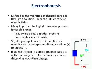
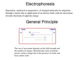
ELECTROPHORESIS • Definition: Electrophoresis is the migration of charged molecules in an electric field. The negative charged particles (anions) moves towards positive charged electrodes (anode). Positively charged particles (cations) moves towards cathod (negatively charged electrode). • Types: • Depending upon the nature of supporting medium a. Agar gel electrophoresis (AGE). b. PAGE, SDS PAGE, QPNC PAGE (Quantitative preparative native continuous PAGE). c. Cellulose acetate electrophoresis. d. Capillary electrophoresis.
Depending upon the mode of technique. a. Slide gel electrophoresis. b. Tube gel electrophoresis. c. Disc electrophoresis. d. Low and high voltage electrophoresis. e. Two dimensional gel electrophoresis Applications of Electrophoresis: • Separating serum proteins for diagnostic purpose. • Haemoglobin separation. • Lipoprotein separation and identification. • Isoenzyme separation and their analysis. • Nucleic acid studies. • Determination of molecular weight of the proteins.
FACTORS AFFECTING ELECTROPHORESIS • The electric field: • Voltage - V ∞ M • Current - C ∞ M • Resistance- R 1/∞ M • The sample: • Charge - C ∞ M • Size - S 1/∞ M • Shape - Molecules of similar size but different shape such as fibrus and globular proteins exhibit diffeent migration characteristics. Because of the differential effect of frictional and electrophoretic force.
III. The buffers: This determines and stabilizes the pH of the supporting medium and hence affects the migration rate of compound in a number of ways. • Composition: The buffer should be such that it does not binds with the compounds to be separated as this may alters the rate of migration. Therefore barbitone buffer is always preferred for the separation of proteins or lipoproteins. • Concentration: As the ionic strength of the buffThe er increases the proportion of current carried by the buffer will increase and the share of the current carried by the sample will decrease thus slowing down the rate of migration. • pH: For organic compounds pH determines the extent of ionization and therefore degree and direction of migration are pH dependent. • The supporting medium: The composition of supporting medium may cause adsorption, electro osmosis and molecular sieving. Which may influence the rate of migration of compounds. The commonly using supporting medium in the laboratory are agarose, polyacrylamide and cellulose acetate membrane.
Types of buffers used in electrophoresis • Tris buffer. • Glycine buffer. • Sodium barbituric acid. • TAE buffer (Tris acidic acid EDTA).
Types of Stains • For serum proteins – Amido block - Coomassie brilliant blue • For isoenzymes - Nitro tetra zolium blue • For lipoprotein zones- Fat red 7B - Oil red O - Sudan block B • For DNA fragments - Ethidium bromide • For CSA proteins - Silver nitrate
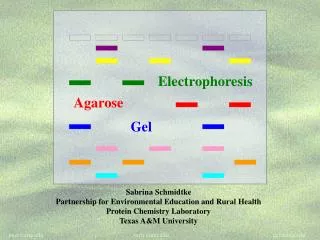
What is needed? • Agarose - a polysaccharide made from seaweed. Agarose is dissolved in buffer and heated, then cools to a gelatinous solid with a network of crosslinked molecules • Some gels are made with acrylamide if sharper bands are required
Buffer - in this case TBE • The buffer provides ions in solution to ensure electrical conductivity. • Not only is the agarose dissolved in buffer, but the gel slab is submerged (submarine gel) in buffer after hardening
Also needed are a power supply and a gel chamber • Gel chambers come in a variety of models, from commercial through home-made, and a variety of sizes
A gel being run Positive electrode Comb Agarose block DNA loaded in wells in the agarose Buffer Black background To make loading wells easier
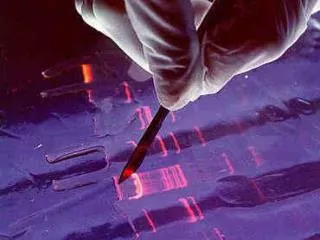
The comb is removed, leaving little ‘wells’ and buffer is poured over the gel to cover it completely • The serum samples are mixed with a dense loading dye so they sink into their wells and can be seen
The serum samples are put in the wells with a micropipette. • Micropipettes have disposable tips and can accurately measure 1/1,000,000 of a litre
Pulsed field gel electrophoresis • Pulsed Field Gel Electrophoresis (commonly abbreviated as PFGE) is a method for separating large DNA molecules, which may be used for genotyping or genetic fingerprinting. • Under normal electrophoresis, large nucleic acid particles (above 30-50 kb) migrate at similar rates, regardless of size. By changing the direction of the electric field frequently, much greater size resolution can be obtained
SDS-PAGE • SDS-PAGE, officially sodium dodecyl sulfate polyacrylamide gel electrophoresis, is a technique used in biochemistry, genetics and molecular biology to separate proteins according to their electrophoretic mobility . • Quantitative preparative native continuous polyacrylamide gel electrophoresis (QPNC-PAGE) is a new method for separating native metalloproteins in complex biological matrices.
NORMAL PATTERN OF SERUM ELECTROPHORESIS 1 origin β2-globulin α2-globulin albumin i β1-globulin α1-globulin γ-globulin
ABNORMAL PATTERN OF SERUM ELECTROPHORESIS MULTIPLE MYELOMA i Extra M-Band is seen Albumin
NEPHROTIC SYNDROME i Β-globulin and α2-gloublin Albumin
AGAMMAGLOBULINEMIA i Absence or decrease of γ-globulin and others normal
LIVER DISEASES origin α2-globulin albumin i α1-globulin γ β-globulin
Characteristic Patterns of Acute-Reaction Proteins Found on Serum Protein Electrophoresis and Associated Conditions or Disorders Increased albumin Dehydration Decreased albumin Chronic cachectic or wasting diseases Chronic infections Hemorrhage, burns, or protein-losing enteropathies Impaired liver function resulting from decreased synthesis of albumin Malnutrition Nephrotic syndrome Pregnancy Increased alpha1 globulins Pregnancy Decreased alpha1 globulins Alpha1-antitrypsin deficiency Increased alpha2 globulins Adrenal insufficiency Adrenocorticosteroid therapy Advanced diabetes mellitus Nephrotic syndrome Decreased alpha2 globulins Malnutrition Megaloblastic anemia Protein-losing enteropathies Severe liver disease Wilson's disease
Increased beta1 or beta2 globulins Biliary cirrhosis Carcinoma (sometimes) Cushing's disease Diabetes mellitus (some cases) Hypothyroidism Iron deficiency anemia Malignant hypertension Nephrosis Polyarteritis nodosa Obstructive jaundice Third-trimester pregnancy Decreased beta1 or beta2 globulins Protein malnutrition Increased gamma globulins Amyloidosis Chronic infections (granulomatous diseases) Chronic lymphocytic leukemia Cirrhosis Hodgkin's disease Malignant lymphoma Multiple myeloma Rheumatoid and collagen diseases (connective tissue disorders) Waldenström's macroglobulinemia Decreased gamma globulins Agammaglobulinemia Hypogammaglobulinemia
Differential Diagnosis of Polyclonal Gammopathy Infections Viral infections, especially hepatitis, human immunodeficiency virus infection, mononucleosis, and varicella Focal or systemic bacterial infections, including endocarditis, osteomyelitis, and bacteremia Tuberculosis Connective tissue diseases Systemic lupus erythematosus Mixed connective tissue Temporal arteritis Rheumatoid arthritis Sarcoid Liver diseases Cirrhosis Ethanol abuse Autoimmune hepatitis Viral-induced hepatitis Primary biliary cirrhosis Primary sclerosing cholangitis Malignancies Solid tumors Ovarian tumors Lung cancer Hepatocellular cancer Renal tumors Gastric tumors Hematologic cancers (see below) Hematologic and lymphoproliferative disorders Lymphoma Leukemia Thalassemia Sickle cell anemia Other inflammatory conditions Gastrointestinal conditions, including ulcerative colitis and Crohn's disease Pulmonary disorders, including bronchiectasis, cystic fibrosis, chronic bronchitis, and pneumonitis Endocrine diseases, including Graves' disease and Hashimoto's thyroiditis