Download

1 / 18
230 likes | 640 Views
Mutations and Gel Electrophoresis. Mutations. “Changes in the DNA sequence that are inherited” Can have a negative consequence, no consequence, or a positive consequence. Silent Mutations. Has no consequence (neither good nor bad) Can happen in 2 ways:

E N D
Mutations • “Changes in the DNA sequence that are inherited” • Can have a negative consequence, no consequence, or a positive consequence
Silent Mutations • Has no consequence (neither good nor bad) • Can happen in 2 ways: • A mutation occurs in an intron, which is spliced out during mRNA processing. • A mutation occurs in the DNA which does not change the amino acid
Silent Mutations - Example Example (No change in aa) 5’ – AUG AAG UUU GGC UAA – 3’ Met Lys Phe Gly Stop 5’ – AUG AAG UUU GGU UAA – 3’ Met Lys Phe Gly Stop Original (normal) mRNA Mutated (Base C changed to U) mRNA But no change in amino acid
Missense Mutations • A change in a nitrogenous base leads to a change in the amino acid produced. 5’ – AUG AAG UUU GGC UAA – 3’ Met Lys Phe Gly Stop 5’ – AUG AAG UUU AGC UAA – 3’ Met Lys Phe Ser Stop Original (normal) mRNA Missense mutation (G base changed to A) = A different amino acid
Nonsense Mutations • A change in a nitrogenous base leads to a premature stop codon 5’ – AUG AAG UUU GGC UAA – 3’ Met Lys Phe Gly Stop 5’ – AUG UAG UUU GGC UAA – 3’ Met Stop Original (normal) mRNA Nonsense mutation (A base changed to U) = Premature stop codon
Classification • Missense and Nonsense mutations arise because of a base pair substitution • In other words, the nitrogenous base itself may change, but the number of nitrogenous bases don’t change
Deletion Mutations • One or more bases are deleted 5’ – AUG AAG UUU GGC UAA – 3’ Met Lys PheGly Stop 5’ – AUG AAG UUG GCU AA – 3’ Met Lys LeuAla Original (normal) mRNA Deletion of the base “U” = The “reading frame” shifts, and new amino acids result
Insertion Mutations • The addition of one or more bases 5’ – AUG AAG UUU GGC UAA – 3’ Met Lys Phe Gly Stop 5’ – AUG UAA GUU UGG CUA – 3’ Met Stop Original (normal) mRNA Insertion of the base “U” = The “reading frame” shifts, and a stop codon results. This frameshift insertion cause a nonsense mutation
Classification • Frameshift mutations occur because of insertions or deletions • All of these mutations (missense, nonsense, insertions, deletions) are called point mutations meaning that they occur only with one base pair
Classification • Another category of mutations involves large segments of DNA • These are called chromosomal mutations TRANSLOCATIONS INVERSIONS
How do mutations arise? • What causes mutations? • List examples • Spontaneously • Mutagenic agents (chemicals that cause mutations, e.g. X rays, UV radiation, cosmic rays, chemicals)
Try It! The following strand of mRNA represents the “normal” (aka wild type) strand. 5’ – AUG GGG UUU AUC CUA UAG – 3’ This strand is hit by UV radiation and turns into: 5’ – AUG GGG UUG AUC CUA UAG – 3’ • Write the amino acid sequences for both strands • What kind of mutation occurred? Be very specific. • What would happen if the “UUG” changed into “UAG”?
Gel Electrophoresis Prep for Tomorrow’s Lab
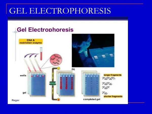
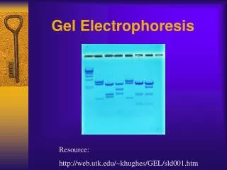
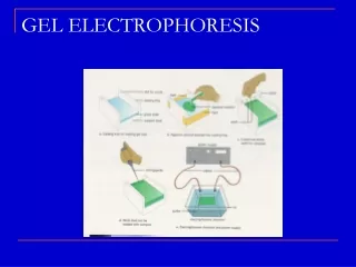
Gel Electrophoresis • Is a procedure used to separate DNA fragments
Gel Electrophoresis – General Steps • Prepare the DNA samples by adding restriction enzymes to them. Restriction enzymes will cut the DNA into smaller fragments. • Add loading dye to your DNA samples. This will allow you to see the DNA as it migrates down the gel. • Pour the agarose gel (with buffer) into your electrophoresis tray.
Gel Electrophoresis – Steps Cont’d 4. Load your DNA into the “wells” using a micropipette. Make sure you write down which sample you are putting into which well (e.g. Well 1 = Crime Scene Suspect, Well 2 = DNA Sample 1, Well 3 = DNA Sample 2, etc.) 5. Plug in your electrodes, and turn on the power to 100V for 30 minutes. 6. Your DNA fragments will separate based on size.
Gel Electrophoresis • Remember that this lab is informal • Try to answer the questions as you perform the lab, it will give you less homework to do afterwards