Download

1 / 19
370 likes | 1.96k Views
Practical Hematology Lab. - LAB 13 -. Hemoglobin Electrophoresis. Electrophoresis. Electrophoresis is a means of separating hemoglobin's .

E N D
Practical Hematology Lab - LAB 13 - Hemoglobin Electrophoresis
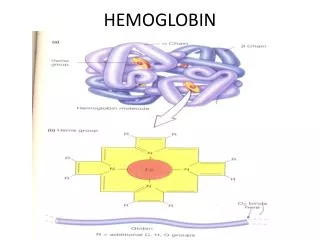
Electrophoresis • Electrophoresis is a means of separating hemoglobin's. • It depends on the migration of the hemoglobin molecules dissolved in a buffer on, or in, a supporting medium when an electric current is passed through them. • Hemoglobin electrophoresis • Is a test that measures the different types of the oxygen-carrying substance (hemoglobin) in the blood. • Hemoglobin electrophoresis is performed to find out abnormal forms of hemoglobin (hemoglobinopathy).
Hemoglobin Electrophoresis • Many different types of hemoglobin (Hb) exist. The most common ones are HbA, HbA2, HbF, HbS, HbC, Hb H, and Hb M. • Healthy adults only have significant levels of HbA and HbA2. • Some people may also have small amounts of HbF (which is the main type of hemoglobin in an unborn baby's body). Certain diseases are associated with high HbF levels (when HbF is more than 2% of the total hemoglobin).
Hemoglobin Electrophoresis • Hb S is an abnormal form of hemoglobin associated with sickle cell anemia. In people with this condition, the red blood cells have a crescent or sickle shape. These misformed cells then break down, or can block small blood vessels. • Hb C is an abnormal form of hemoglobin associated with hemolytic anemia. The symptoms are much milder than they are in sickle cell anemia.
Normal Values • In adults: • Hgb A1 : 95% to 98% • Hgb A2 : 2% to 3% • Hgb F : 0.8% to 2% • Hgb S : 0% • Hgb C : 0% • In infants and children: • Hgb F (newborn) : 50% to 80% • Hgb F (6 months) : 8% • Hgb F (over 6 months) : 1% to 2%
Methods of Electrophoresis 1-Cellulose Acetate At Alkaline pH 2- Citrate Agar Electrophoresis ( acid pH)
1-Cellulose Acetate At Alkaline pH • Cellulose acetate Hb electrophoresis at alkaline pH is the primary screening procedure used to detect variant (abnormal) Hbs, of which there are several hundreds. • The major portion of normal adult Hb is A. In addition, up to 3.5% Hb A2 is normally present, along with less than 2% Hb F. The more common mutant Hbs are S, C, E, D, G, and lepore.
1-Cellulose Acetate At Alkaline pH • When an abnormal Hb is detected on cellulose acetate electrophoresis at an alkaline pH (8.2-8.6) further testing is frequently indicated: test for Hb S, quantitation of Hb A2 and F, and citrate agar gel; acid/alkaline globin chain or neutral pH electrophoresis may also be warranted.
Principle of Cellulose Acetate • In an alkaline pH (8.2-8.6) Hb is a negatively charged molecule and will migrate toward the anode (+). The various Hbs moves at different rates depending on their net negative charge, which in turn is controlled by the composition (amino acids) of the Hb molecule (globin chain).
Principle of Cellulose Acetate • The red cell hemolysate (red blood cell membranes are destroyed to free the Hb molecules for testing) is placed in a cellulose acetate membrane, which is positioned in an electrophoresis tray with the inoculated hemolysate near the cathode (-).
Principle of Cellulose Acetate • One end of the cellulose acetate strip is immersed in the buffer (pH 8.2-8.6) on the cathode side and the other end is placed in the buffer on the anode (+) side. An electric current of specific voltage is allowed to run for a timed period. • During electrophoresis, the Hb molecules migrate toward the anode because of their negative charge. The difference in the net charge of the Hb molecule determines its mobility and manifests its self by the speed with which it migrates to the positive pole.
Principle of cellulose acetate • The cellulose acetate membrane is then stained in order to color the proteins (Hbs). By noting the distance each Hb has migrated and comparing this distance with the migration distance of known controls, the types of hemoglobins may be identified. • Example of the fast Hbs are Hb Bart’s and the two fastest variants Hb H and I, while Hb C is the slowest common Hb.
2- Citrate Agar Electrophoresis ( acid pH) • Citrate agar separates Hb fractions that migrate together on cellulose acetate agar. • All Hb specimens that show an abnormal electrophoretic pattern in alkaline media (cellulose acetate agar) should undergo electrophoresis on an acid citrate agar.
2- Citrate Agar Electrophoresis ( acid pH) • Citrate agar electrophoresis is used to confirm variant Hbs and further differentiates Hb S from Hb D and G, and Hb C from Hb E, O Arab, and CHarlem. . • Theprocedure should not be used as a screening procedure because many abnormal Hbs migrate with Hb A. However, this procedure is the method of choice when examining newborns (cord blood specimens) and infants under 3 months of age for some abnormal Hbs such as S and C because the test is able to detect quantities of Hb not easily seen by other techniques.
C s A F D G A2 E O