Download
1 / 17
170 likes | 283 Views
Antigen Processing and Presentation, Self MHC Restriction, Role of Thymus, and Superantigens. What Does The B Cell Immunoglobulin (Ig) Receptor Recognize?. Proteins (conformational determinants, denatured or proteolyzed determinants) Nucleic acids Polysaccharides Some lipids

E N D
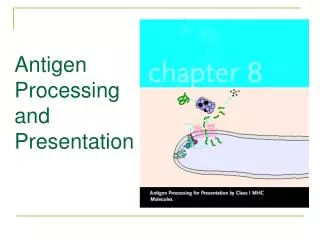
Antigen Processing and Presentation, Self MHC Restriction, Role of Thymus, and Superantigens
What Does The B Cell Immunoglobulin (Ig) Receptor Recognize? • Proteins (conformational determinants, denatured or proteolyzed determinants) • Nucleic acids • Polysaccharides • Some lipids • Small chemicals (haptens)
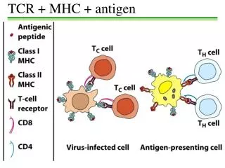
What Does the αβ T Cell Receptor (TCR) Recognize? • Only fragments of proteins (peptides) associated with MHC molecules on surface of cells • Helper T cells (Th) recognize peptide associated with MHC class II molecules • Cytotoxic T cells (Tc) recognize peptide associated with MHC class I molecules
Experimental Basis for Antigen Processing and Presentation • Pulse macrophages with a protein antigen short time, wash cells, fix immediately, mix with T cells and measure proliferation • NO PROLIFERATION OF T CELLS • Pulse macrophages with same protein antigen short time, wash cells, fix after 4-5 hours, mix with T cells and measure proliferation • PROLIFERATION OF T CELLS
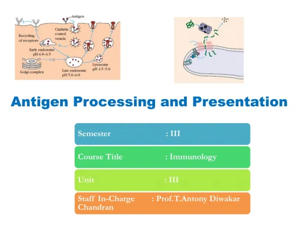
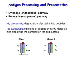
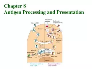
Antigen Processing and Presentation • Fragmentation of protein into peptides • Association of peptide with an MHC molecule • Transport to cell surface for expression • Different cellular pathways for association of peptide with MHC class I and class II molecules
Viral protein is made oncytoplasmic ribosomes Plasma membrane Class I MHC Pathway Peptide is presented by MHC-I to CD8 cytotoxic T cell Globular viral protein - intact Peptide passes with MHC from Golgi body to surface Proteasome degrades protein to peptides rER Peptide associates with MHC-I complex Peptide transporter protein moves peptide into ER MHC class I alpha and beta proteins are made on the rER Peptide with MHC goes to Golgi body Golgi body
Peptide MHC-II complex is presented to CD4 helper T cell Globular protein CD4 helper T cell Endosome Endosome fuses with plasma membrane Endocytosis Fusion of endosome and exocytic vesicle Immunodominant peptide binds to class II MHC Lysosome Exocytic vesicle fuses with endosome releasing Ii from αβ dimer Protein is processed to peptides in endosome or lysosome Golgi body Class II MHC Synthesis 3 chains: α,β and Ii α β Ii Endoplasmic reticulum Class II MHC Pathway
Points Concerning Antigen Processing and Presentation 1. Location of pathogen • viruses in cytosol, MHC class I pathway, Tc response • extracellular bacteria, MHC class II pathway, Th2 response, Ab formation • intracellular bacteria, MHC class II pathway, Th1 response
Points Concerning Antigen Processing and Presentation • Peptides derived from both self and non-self proteins can associate with MHC class I and class II molecules. • Chemical nature of MHC groove determines which peptides it will bind.
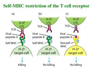
Self MHC Restriction • T cells recognize foreign antigen associated with self MHC • No value for individual to have T cells that recognize foreign antigen associated with foreign MHC • Self MHC restriction occurs in thymus
Mouse Strain A or B Mouse Strain A or B Immunize with antigen Period of 7 days Peritoneal macrophage Antigen T cells isolated from lymph nodes Macrophage presents antigen Anti X-induced MacrophageT-cellproliferation Strain A A yes Strain A B no Strain B B yes Strain B A no T cell primed with antigen Assay DNA synthesis after 48 hr Macrophages and T cells are co-cultivated Th Cell Self MHC Restriction
Mouse Strain A or B Mouse Strain A or B Immunize with virus PurifyFibroblasts Period of 7 days Isolate spleen Infect fibroblasts with virus and radiolabel with 51Cr Virus-specific cytotoxic T cells (CTLs) isolated from spleen 51Cr labeled fibroblast presents antigen Spleen Fibroblast 51Cr release CTLs (target cells) from fibroblasts immunized infected (lysis) withwith Strain A A yes Strain A B no Strain B B yes Strain B A no Virus-specificCTLs Assay 51Cr release CTLs and fibroblasts are co-cultivated Tc Cell Self MHC Restriction
Process of Self MHC Restriction in Thymus • T cells with TCR recognizing self MHC molecules are retained – “positive selection” • Retained T cells with TCR recognizing self peptide associated with self MHC are eliminated – “negative selection” • Self MHC-restricted T cells are released
Medulla vessel 4 - 8 + TCR 4 + 8 - TCR Self MHC Restriction in the Thymus 4 low 8 low 4 - 8 low Sub-capsular region Productive TCR rearrangement Non-productive TCR rearrangement APOPTOSIS 4 + 8 + TCR Not recognise self MHC Recognise self MHC macrophage 4 + 8 + TCR TCR recognisesself antigens Cortex TCR does not recognise self antigens Negative selection Cortico-medullary region 4 - 8 - 4 + 8 + TCR
Superantigens • Proteins produced by pathogens • Not processed by antigen presenting cells • Intact protein binds to variable region of β chain on TCR of T cells and to MHC class II on antigen presenting cells (APC) • Large numbers of activated T cells release cytokines having pathological effects
Conventional Antigen Antigen presenting cell α2 β2 α2 β2 CHO CHO MHC Class II α1 β1 α1 β1 CHO CHO CHO CHO Super antigen Antigen αV αV βV βV CHO CHO CHO CHO T cell receptor αC βC αC βC CHO CHO CHO CHO T lymphocyte Superantigen