Download
1 / 34
500 likes | 1.48k Views
Antigen Processing and Presentation. Recognition of foreign antigen by a T cell requires that peptides derived from the antigen be displayed within the cleft of an MHC molecule on the membrane of a cell. Exogenous Ag. Endogenous Ag.

E N D
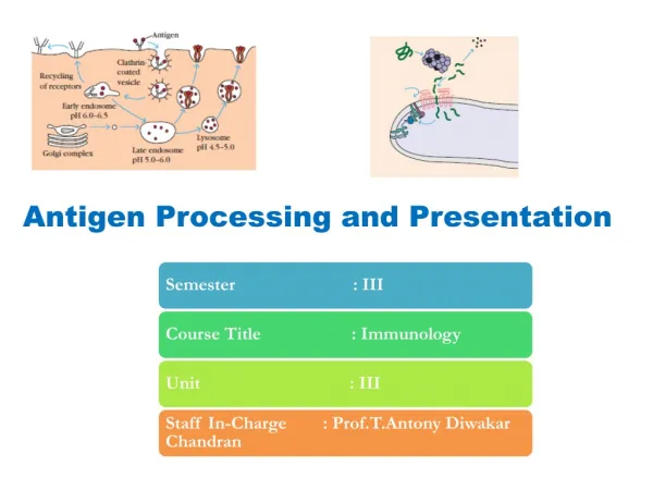
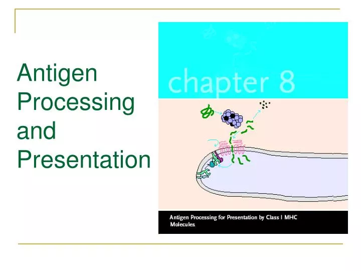
Antigen Processing and Presentation
Recognition of foreign antigen by a T cell requires that peptides derived from the antigen be displayed within the cleft of an MHC molecule on the membrane of a cell.
Antigen processing: The formation of peptide-MHC complexes requires that a protein antigen be degraded into peptides by a sequence of events. Antigen presentation: The degraded peptides then associate with MHC molecules within cell interior, and the peptide-MHC complexes are transported to the membrane,where they are displayed.
Self-MHC restriction :Both CD4+ and CD8+ T cells can recognize antigen only when it is presented with a self-MHC molecule , an attribute called self-MHC restriction.
Self-MHC restriction was first discovered in experiments in which T cells from one inbred strain were mixed with macrophages, B cells, or virus-infected cells from another inbred strain. In each experiment, T cells responded to the antigen only if they share MHC alleles with the other cells in mixture. For example: A. researchers showed that antigen-specific proliferation of TH cells occurred only in response to antigen presented by macrophages of the same MHC haplotype.
Results Experimental demonstration of self-MHC restriction of TH
LCM: Lymphocytic choriomeningitis (淋巴细胞性脉络丛脑膜炎) Cr: chromium(铬) Antigen recognition by Tc cells exhibits HMC restriction
2.Role of Antigen-presenting Cells The macrophage presents processed antigen combined with class II MHC molecules to the T cells.
1) Processing of Antigen Is Required for Recognition by T Cells a.When APCs are fixed before exposure to antigen, they are unable to activate TH cells. b.In contrast, APCs fixed at least 1 h after antigen exposure can activate TH cells. c. When APCs are fixed before antigen exposure and incubated with peptide digests of the antigen,they also can activate TH cells.
2) Most Cells Can Present Antigen with Class I MHC; Presentation with Class II MHC Is Restricted to APCs Since all cells expression either class I and II MHC molecules can present to T cells, strictly speaking they all could be designated as APCs. However,by convention, cells that display peptides associated with class I MHC molecules to CD8+ Tc cells are referred to as target cells. Cells that display peptides associated with class II MHC molecules to CD4 + Th cells are called antigen-presenting cells.
Dendritic cells are the most effective of the antigen- presenting cells.Because these cells constitutively express a high level of class II MHC molecules and co-stimulatory activity, they can activate naïve Th cells. Macrophages must be activated by phagocytosis of microorganisms before they express class II MHC molecules or co-stimulatory B7 membrane molecules
B cells constitutively express class II MHC molecules but must be activated before they express the co-stimulatory B7 molecules.
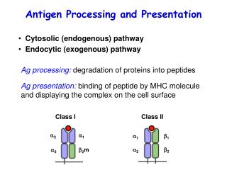
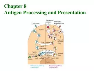
3. Evidence for Two Processing and Presentation Pathways The immune system uses two different pathways to eliminateintracellular and extracellular antigens. Endogenous antigens(those generated within the cell) are processed in the cytosolicpathwayand presented on the membrane with class IMHC molecules; exogenous antigens (those taken up by endocytosis)are processed in the endocytic pathway and presentedon the membrane with class II MHC molecules.
Overview of cytosolic and endocytic pathways for processing antigen. The proteasome complex contains enzymes that cleave peptide bonds, converting proteins into peptides. The antigenic peptides from proteasome cleavage and those from endocytic compartments associate with class I or class II MHC molecules, and the peptide-MHC complexes are then transported to the cell membrane. TAP (transporter of antigenic peptides) transports the peptides to the endoplasmic reticulum内质网.
4. Endogenous Antigens: The Cytosolic Pathway Endogenous antigens, such as those produced by a virus replication within a cell, are degraded within the cytoplasm into peptides that can associate with class I MHC molecules.
1) Peptides for Presentation Are Generated by Protease Complexes Called Proteasomes(蛋白酶体) 赖氨酸 泛素
2) Peptide transport from the cytosol to rough endoplasmic reticulum(RER,粗面内质网) TAP-Transporter associated with antigen processing TAP anchored in the membrane of RER. The two chains are encoded by TAP1 and TAP2.
1.Within the RER membrane,newly synthesized class I α chain associates with calnexin until β2-microglobulin binds to the α chain. 2.The class I α chain- β2-microglobulin heterodimer then binds to calreticulin and the Tapasin. 3.When a peptide delivered by TAP is bound to the class I molecule, the folding of MHC class I is complete and it is released from the RER and transported through the Golgi to the surface of cells. TAP-associated protein 钙网蛋白 钙联蛋白 Generation of antigenic peptide-class I MHC complexes in cytosolic pathway
3) Peptides Assemble with Class I MHC Aided by Chaperone Molecules Assembly and stabilization of class I MHC molecules. Newly formed class I chains associate with calnexin, a molecular chaperone伴护, in the RER membrane. Subsequent binding toβ 2-microglobulin releases calnexin and allows binding to the chaperonin calreticulin and to tapasin, which is associated with the peptide transporter TAP. This association promotes binding of an antigenic peptide, which stabilizes the class I molecule–peptide complex, allowing its release from the RER.
Separate antigen-presenting pathways are utilized for endogenous (green) and exogenous (red) antigens. The mode of antigen entry into cells and the site of antigen processing determine whether antigenic peptides associate with class I MHC molecules in the rough endoplasmic reticulum or with class II molecules in endocytic compartments.
5. Exogenous Antigens: The Endocytic Pathway Antigen-presenting cells can internalize antigen by phagocytosis, endocytosis, or both. Macrophages internalize antigen by both processes, whereas most other APCs are not phagocytic or are poorly phagocytic and therefore internalize exogenous antigen only by endocytosis (either receptor-mediated endocytosis or pinocytosis). B cells, for example, internalize antigen very effectively by receptor-mediated endocytosis using antigen-specific membrane antibody as the receptor.
1) Peptides Are Generated from Internalized Molecules in Endocytic Vesicles 网格蛋白 Generation of antigenic peptides in the endocytic processing pathway
2) The Invariant Chain Guides Transport of Class II MHC Molecules to Endocytic Vesicles
3) Peptides Assemble with Class II MHC Molecules by Displacing CLIP CLIP: class II–associated invariant chain peptide)
SUMMARY • T-cells recognize antigen displayed within the cleft of a self-MHC molecule on the membrane of a cell. • 2. In general, CD4 TH cells recognize antigen with class II MHC molecules on antigen-processing cells. • 3. CD8 TC cells recognize antigen with class I MHC molecules on target cells.
4.Complexes between antigenic peptides and MHC molecules are formed by degradation of a protein antigen in one of two different antigen-processing pathways. 5. Endogenous antigens are degraded into peptides within the cytosol by proteasomes and assemble with class I molecules in the RER.
6. Exogenous antigens are internalized and degraded within the acidic endocytic compartments and subsequently pair with class II molecules. 7. Presentation of nonpeptide (lipid and glycolipid) antigens derived from bacteria involves the class I–like CD1 molecules.