Download
1 / 39
490 likes | 927 Views
Antigen presenting cells and antigen presentation. Contents. Antigen presenting cells Antigen presentation. Antigen presenting cell, APC. A variety of cell types which carry antigen in a form that can stimulate lymphocytes. It is also termed accessory cell. professional APC:M Φ ,DC,B

E N D
Antigen presenting cells and antigen presentation
Contents • Antigen presenting cells • Antigen presentation
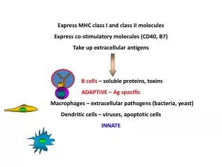
Antigen presenting cell, APC A variety of cell types which carry antigen in a form that can stimulate lymphocytes. It is also termed accessory cell. professional APC:MΦ,DC,B non-professional APC
The 3 types of professional APCs Constitutively express a high level of MHC II and the co-stimulatory protein,B7. the most effective APC must be activated by the process of phagocytosis before expressing class II MHC and B7. Constitutively express class II MHC but must be activated to produce B7.
I. Dendritic cells (DC) • Dendritic cells were first described by Paul Langerhans (Langerhans cells) in the late nineteenth century. It wasn't until 1973, however, that the term "dendritic cells" was coined by Ralph M. Steinman and Zanvil A. Cohn. In 2007 Steinman was awarded the Albert Lasker Award for Basic Medical Research for his discovery.
Marks • Mouse 33D1,NLDC145 • Rat OX62 • Human CD1a,CD11c,CD83 • Other marks:MHCⅡ,CD80,CD86
HSC Myeloid progenitor GM-CSF TNF-a IL-4 Sources of DC Lymphoid progenitor DC Mo PMN DC DC macrophage
Dendritic Cell Maturation LPS IL-1,TNFα
B Menu F
Functions of DC: antigen presentation, immune activation, immune tolerance
Antigen presenting cells mononuclear phagocyte system, MPS 1.Differentiation and distribution 2. Surface markers 3. function
Differentiation and distribution of MPS Bone marrow Blood tissues HSC monocyte macrophage Myeloid progenitor Pre-monocyte monocyte
Mononuclear phagocyte system, MPS 1. Surface markers: MHC-I/Ⅱmolecules CKR: M-CSFR CAM: LFA-1,ICAM-1,B7,CD40 FcR, CR1/3/4 2. secretion: -enzymes: lysosome , myeloperoxidase -cytokines (IL-1,TNF,IL-12) -complement:C1~C9, Bf -coagulation factor, PG, LTs, ACTH, etc.
Biological functions of MPS • Phagocytosis • antitumor:indirect or direct killing, ADCC; • participating in immune response: -Ag presenting, providing the second signal; -CMI • participating in immune regulation: -positive regulation:secrete IL-1/12,TNF-α -negative regulation:PG, TGF-β • Mediating inflammation: phagocytosis, secrete inflammation medium
Antigen presenting cells B cells
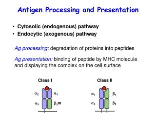
Ag presentation • Definition • APC • Ag presentation -MHC class I molecule pathway -MHC class II molecule pathway
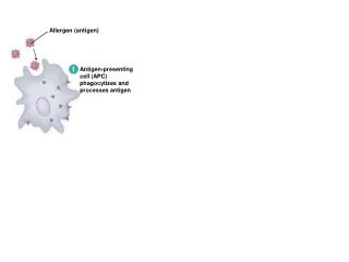
Antigen capturing • Phagocytosis • Pinocytosis • Receptor-mediated endocytosis
Antigen processing The conversion of an antigen into a form in which it can be recognized by lymphocytes.
Antigen presentation The process by which certain cells in the body (APC) express antigen peptide-MHC molecule complex on their cell surface in a form recognize by lymphocytes.
Recourses of antigens * exogenous antigen * endogenous antigen
Y The site of pathogen replication or mechanism of antigen uptake determines the antigen processing pathway used EXTRACELLULAR OR ENDOSOMAL REPLICATION Y Vesicular Compartment Contiguous with extracellular fluid Exogenous processing (Streptococcal, Mycobacterial antigens) INTRACELLULAR REPLICATION Cytosolic compartment Endogenous processing (Viral antigens) Distinct mechanisms of antigen generation are used to raise T cells suited to the elimination of endogenous or exogenous pathogens
The pathway of MHC I -associated endogenous Ag presentation endogenous antigen(such as virus Ag, tumor Ag) cytoplasm degraded by proteasome antigen peptide(8-13 AA) transported to endoplasmic reticulum by TAP Peptide/MHC-I molecule complex to surface of APC submit to CD8+T
Degradation in the proteasome Cytoplasmic cellular proteins, including non-self proteins are degraded continuously by a multicatalytic protease of 28 subunits The components of the proteasome include MECL-1, LMP2, LMP7 These components are induced by IFN- and replace constitutive components to confer proteolytic properties. LMP2 & 7 encoded in the MHC Proteasome cleaves proteins after hydrophobic and basic amino acids and releases peptides into the cytoplasm
Hydrophobic transmembrane domain Lumen of ER Lumen of ER Peptide Peptide Peptide Peptide Peptide Peptide Peptide Peptide Peptide Peptide Peptide ER membrane ER membrane TAP-1 TAP-1 TAP-1 TAP-1 TAP-1 TAP-1 TAP-1 TAP-1 TAP-1 TAP-1 TAP-1 TAP-2 TAP-2 TAP-2 TAP-2 TAP-2 TAP-2 TAP-2 TAP-2 TAP-2 TAP-2 TAP-2 Cytosol Cytosol ATP-binding cassette (ABC) domain Peptide antigens from proteasome Transporters associated with antigen processing (TAP1 & 2) Transporter has preference for >8 amino acid peptides with hydrophobic C termini.
Peptide Peptide Peptide Peptide Peptide Peptide Peptide Peptide Peptide Peptide Peptide TAP-1 TAP-1 TAP-1 TAP-1 TAP-1 TAP-1 TAP-2 TAP-2 TAP-2 TAP-2 TAP-2 TAP-2 TAP-2 TAP-2 TAP-2 TAP-2 TAP-2 TAP-1 TAP-1 TAP-1 TAP-1 TAP-1 Endoplasmic reticulum Maturation and loading of MHC class I B2-M binds and stabilises floppy MHC Tapasin, calreticulin, TAP 1 & 2 form a complex with the floppy MHC Calnexin binds to nascent class I chain until 2-M binds Cytoplasmic peptides are loaded onto the MHC molecule and the structure becomes compact
B Menu F
B Menu F
The pathway of MHC II -associated exogenous Ag presentation Exogenous antigen newly synthesised MHC class II molecule (in the endoplasmic reticulum) early endosome li binds in the groove of MHC class II molecule lysosomeproteaseMIIC late endosomeli degrade protease Degrade into 1318AA peptide + MHC class II molecule Ag peptide/MHC class II molecule complex transport to the surface of APC, recognized by CD4+T Phagocytosis, pinocytosis, FcR-phagocytosis
Y Y Y Uptake of exogenous antigens Membrane Ig receptor mediated uptake Y Phagocytosis Complement receptor mediated phagocytosis Pinocytosis opsonization Fc receptor mediated phagocytosis
Uptake Endosomes Increase in acidity To lysosomes Exogenous pathway Protein antigens In endosome Cathepsin B, D and L proteases are activated by the decrease in pH Proteases produce ~24 amino acid long peptides from antigens Drugs that raise the pH of endosomes inhibit antigen processing
The functions of Ii: • involve in the assembling and folding of MHC class II molecule; • Block the groove of MHC class II molecule; • Lead the assembled class II molecule to MⅡC. CLIP:class II-associated invariant chain peptide
B Menu F
B Menu F
Cross-presentation • Class I MHC molecules present exogenous Ags to CD8+ T cells • Cross-presentation of Ags by DC plays an important role in anti-viral infection and anti-tumor immunity.