Download
1 / 19
380 likes | 1.73k Views
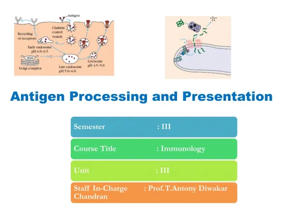
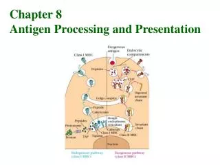
Class I. Class II. 2. 1. 1. 1. 2. 2. 3. 2 m. Antigen Processing and Presentation. Cytosolic (endogenous) pathway Endocytic (exogenous) pathway. Ag processing: degradation of proteins into peptides.

E N D
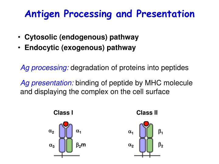
Class I Class II 2 1 1 1 2 2 3 2m Antigen Processing and Presentation • Cytosolic (endogenous) pathway • Endocytic (exogenous) pathway Ag processing: degradation of proteins into peptides Ag presentation: binding of peptide by MHC molecule and displaying the complex on the cell surface
Degraded in Peptides bind to Presented to Intracellular pathogens Cytosol (endogenous) Class I CD8 T cells Two compartments of the cell Cytosol: continuous with nucleus Vesicular system (ER, golgi, endosomes, lysosomes): continuous with extracellular fluid Extracellular pathogens Endocytic vesicles (exogenous) Class II CD4 T cells
Plasma membrane nucleus ER Golgi vesicle Cytosolic pathway Site of peptide generation Site of membrane protein synthesis Transport of peptides into ER Loading of peptide onto nascent class I molecules in ER Display the complex on the cell surface
Plasma membrane nucleus ER Golgi vesicle Cytosolic pathway
X • US6 blocks TAP • US3 prevents class I from egress from ER • US18 mimics HLA-E Pathogen exploitation Plasma membrane nucleus ER Golgi vesicle • Human Cytomegalovirus (HCMV): • US2 and US11 bind and remove nascent class I a chain from ER
Y Y Y Y Endocytic pathway Site of peptide generation Site of MHC class II synthesis Loading of peptide into class II molecules Surface expression Plasma membrane
Y Y Plasma membrane Y Y Endocytic pathway
Influenza A Virus • Segmented RNA virus • 8 RNAs • 10 proteins • 15 HA • 8 NA • H5N1
Antigenic Drift: a series of mutations that occur over time and cause a gradual evolution of the virus Seasonal Influenza Epidemics cRNA (+) • RNA polymerase • No proof-reading • No repair • High mutation rate • 1.5 x 10⁻⁵/nt/cycle RNA pol Virion vRNA (-) RNA pol mRNA (+) Virion RNA Complementary RNA Messenger RNA protein
Antigenic Shift: an abrupt change in the HA and/or the NA proteins resulting in a new subtype of the virus Influenza Pandemics Cell Co-infection Reassortment
Interferons inhibit protein synthesis and therefore virus replication
Role of T and B cells in responses to influenza virus infection CD8 CD4 B Clearance (days) % Survival + + + 7-10 100 - + + 10-14 100 + - + 10-14 90 + + - 10-14 35-85 - - + >20 0 - + - >20 0 + - - >14 20 - - - >20 0
Host-Influenza Virus Interaction Host Influenza virus 5’ triphosphate 5’ cap TLR7, RIG-I dsRNA, NS1 APOBEC NK cells Antibody HA and NA (antigenic shift) CTL Epitope change (antigenic drift)
Principles of adaptive immunity • TCR recognition • Antigen presentation and processing • Host defense against viruses