Download
1 / 57
700 likes | 1.15k Views
Histology of the Female Reproductive System. Reproductive function in the male primarily involves a single organ (testis) and the process is continuous. Reproductive function in the female requires three organs (ovary, uterus and breast) and the process involves interrelated cycles. Oviduct.

E N D
Histology of the Female Reproductive System
Reproductive function in the male primarily involves a single organ (testis) and the process is continuous. Reproductive function in the female requires three organs (ovary, uterus and breast) and the process involves interrelated cycles.
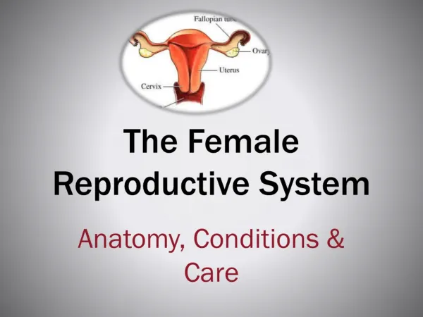
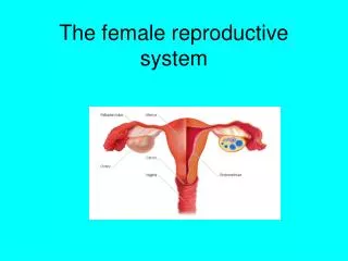
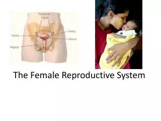
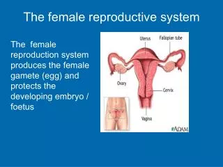
Oviduct Uterus Ovary Cervix Vagina Oviduct Uterus Ovary Cervix Organs Ovaries – paired organs Oviducts – paired organs Uterus (includes cervix) Vagina
Ovarian cycle Uterine (menstrual) cycle Ovarian and uterine cycles Ovulation Progesterone Estrogen Estrogen
Ovarian cycle Uterine (menstrual) cycle Ovarian and uterine cycles Estrogen
The ovarian cycle (28 days)
The ovarian cycle (28 days) - Follicular phase (days 1-13): Follicle differentiation, estrogen secretion. (Uterine/menstrual cycle: Proliferative phase)
The ovarian cycle (28 days) oocyte - Follicular phase (days 1-13): Follicle differentiation, estrogen secretion. - Ovulation (day 14): Graafian follicle ruptures, releasing the oocyte.
The ovarian cycle (28 days) - Follicular phase (days 1-13): Follicle differentiation, estrogen secretion. - Ovulation (day 14): Graafian follicle ruptures, releasing secondary oocyte. - Luteal phase (days 15-28): Corpus luteum forms, progesterone and estrogen secretion. (Uterine/menstrual cycle: Secretory phase)
Ovary Tunica albuginea Cortex Germinal epithelium and tunica albuginea Follicles – functional during first half of the cycle; site of oocyte maturation and estrogen production Corpus luteum – present during second half of the cycle produces progesterone Degenerating structures: Atretic follicles Corpus albicans Medulla CT, blood vessels, nerves
Ovarian follicles Primordial Primary Unilaminar Multilaminar Secondary Graafian/Mature
Primordial follicles Contains a primary oocyte Follicular cells form a simple squamous epithelium with a basement membrane; their free surfaces contact the oocyte The only follicle present until puberty
Primary follicles Follicles begin to develop at puberty (5-15 per cycle) primarily in response to follicle stimulating hormone (FSH) secreted by gonadotrope cells in the pituitary gland Primary unilaminar follicle
Primary oocyte Primary follicles Theca folliculi Zona pellucida Primary multilaminar follicle Zona pellucida: Extracellular shell surrounding and formed by the oocyte Glycosaminoglycans and glycoprotein Theca folliculi: Stromal (CT) cells outside the basement membrane of the follicular cells, differentiate to form theca interna and theca externa
Zona pellucida Secondary follicles Primary oocyte Antral spaces Cumulus oophorus Follicle cells Granulosal cells Stratum granulosum Antrum Zona pellucida
Cumulus oophorus Stratum granulosum Secondary follicles Antrum Theca folliculi
Theca folliculi – Cells surrounding the follicle Basement membrane Theca interna Theca externa Antral space Antral space Stratum granulosum Theca folliculi Theca interna cells respond to LH and secrete androgens which are modified (aromatized) by stratum granulosum cells to form estrogen
Mature/Graafian follicle In a normal cycle, only a single secondary follicle progresses to the mature follicle stage Present only during the day preceding ovulation (~ day 14) Contains the oocyte which will be ovulated
Stigma Mature/Graafian follicle Antral space accumulates fluid, follicle size increases Oocyte and surrounding cumulus oophorus cells (corona radiata) detach from the granulosa layer and lie free in the antral space Follicle enlarges (~1-2 cm) bulges from surface forming a stigma Corona radiata
Oocyte surrounded by corona radiata Ovulation Day 14 of ovarian cycle Stimulated by luteinizing hormone (LH) surge Graafian follicle ruptures at the stigma, releasing the oocyte, corona radiata of the cumulus oophorus, follicular liquid and blood (mittelschmerz = middle pain) Oocyte and the surrounding cumulus cells are captured by the oviduct transported toward the uterus. The follicle wall, stratum granulosum, and theca interna are converted into the corpus luteum.
First observation of human ovulation Lousse JC, Donnez J. Fertil Steril. 2008. 90:833. Laparoscopic observation of spontaneous human ovulation.
Corpus luteum Functional during the second half of the ovarian cycle Secretes progesterone & estrogen Composed of: - Granulosa lutein cells - form from cells of the granulosa layer; major component - Theca lutein cells - form from theca interna cells; minor component
Corpus luteum Ovulation Granulosal lutein cells Thecal lutein cells
In a non-pregnant cycle, the corpus luteum is functional for about 12 days after ovulation after which it degenerates as the corpus albicans If pregnancy occurs, corpus luteum is maintained by hormones secreted by the developing embryo and the placenta Corpus albicans
Corpus albicans Degenerating stage of the corpus luteum Mass of white scar tissue composed of collagen and scattered fibroblasts Gradually regresses
Primary follicle Secondary follicle Degenerating stage of follicles (atresia) Can occur at any stage of follicular development Atretic follicles Antral space Oocyte Glassy membrane Remnants of the basement membrane (glassy membrane)
Polycystic ovary syndrome (PCOS) Most common hormonal disorder among women of reproductive age Leading cause of infertility Unknown cause Enlarged ovaries with a thick, scarred capsule; and multiple cysts (unruptured follicles). Follicles are fluid-filled; contain immature oocytes. Lack of regular ovulatory and menstrual cycles and excessive mounts of androgenic (masculinizing) hormones. Abnormally high ratio of LH to FSH ratio and high insulin levels
Primary multilaminar follicle Primary unilaminar follicle Secondary follicles Primordial follicle Corpus albicans Corpus luteum Graafian follicle Ovulation The ovarian cycle - Follicular phase (days 1-13): Follicle differentiation, estrogen secretion - Ovulation (day 14): Graafian follicle ruptures, releasing secondary oocyte - Luteal phase (days 15-28): Corpus luteum forms, progesterone and estrogen secretion
Oogenesis LH surge 24 hrs before ovulation
Pro- nuclei Polar bodies Sperm penetration Secondary oocyte First polar body LH surge (Graafian follicle)
Oogenesis ~1000 oogonia migrate from the yolk sac Expand to 5-7 million develop in the ovary Numbers decline to <1 million primary oocytes at birth ~400 mature and are ovulated Number of mature ova equals the number of fertilizations
Oviduct Site of fertilization Intramural * Ampulla Isthmus Infundibulum with fimbria Transports secondary oocyte, sperm, embryo Site of fertilization Provides nutrients
Ampulla Intramural Number of cilia decrease Number of secretory cells increase Mucosal folds decrease Oviduct Simple columnar ciliated epithelium with secretory (peg) cells Oviducts reach maximal development around ovulation (day 14) due to high estrogen levels Peg cells
Fundus Body Isthmus Cervix Vagina Uterus - Regions
Endometrium Myometrium Perimetrium Uterus (fundus and body)
Endometrium Endometrium Simple columnar epithelium Lamina propria (endometrial stroma) Simple (branched) tubular glands Functional zone(stratum functionalis) Highly proliferative Proliferates in response to ovarian hormones Sloughed during menstruation Basal zone(stratum basalis) Retained following menstruation Regenerates stratum functionalis Myometrium
Blood supply Uterine arteries Arcuate arteries Radial arteries Basal (straight) arteries Supply stratum basalis Spiral arteries Supply the stratum functionalis
Ovarian cycle Uterine (menstrual) cycle Menstrual Days 1-4 Proliferative Days 5-14 Secretory Days 15-26 Premenstrual Days 26-28 Ovulation Uterine cycle (Menstrual cycle)
Uterine cycle (Menstrual cycle) The days of the menstrual cycle are numbered according to the beginning of menses, however this event actually represents the end of the process. The new cycle actually begins around day 5 with the proliferative phase during which the sloughed endometrium is regrown. DayPhase 5-14 Proliferative phase 15-26 Secretory phase 26-28 Ischemic phase of secretory phase 1-4 Menstrual phase
Stratum functionalis Stratum basalis Proliferative phase (days 5-14) Ovarian follicles are growing and secreting estrogen The functional zone regenerates from the basal zone Rapid proliferation of epithelial and stromal cells Glands are straight, not secreting
Prol. Secretory early late Uterine glands Secretory phase (Days 15-26) Ovulation has occurred; corpus luteum secretes progesterone (and estrogen) Endometrium increases in thickness Glands enlarge, become tortuous, coiled and fill with secretory products Spiral arteries lengthen and become highly coiled
Secretory phase (Days 15-26) Predecidual cells Stromal cells enlarge, accumulate glycogen and lipid become pre-decidual cells Pre-decidual cells form the decidua of the maternal placenta and provide initial nutrition for the embryo If pregnancy does not occur, these cells are sloughed with the menses
Pre-menstrual phase (Ischemic portion of secretory phase) (Days 26-28) Estrogen and progesterone production from corpus luteum decreases Cycles of compression and reopening of spiral arteries leads to ischemia, necrosis and extravasation of blood in the stratum functionalis
Menstrual phase (Days 1-4) Functional zone becomes necrotic and is sloughed as menses Basal zone remains, regenerates functional zone
Menstrual cycle Ovulation Proliferative Secretory Menstrual
Ovarian and uterine cycles Estrogen Progesterone and estrogen From follicles From corpus luteum Days 1-14 of cycle Days 15-26 of cycle
....if fertilization occurs Blastocyst implants about day 20-21 when the endometrium is thickest, glands are secretory and spiral arteries well developed The blastocyst secretes a hormone (HCG, human chorionic gonadotrophin) that stimulates and maintains the corpus luteum, thus continuing progesterone and estrogen secretion and prohibiting pre-menstrual and menstrual phases
Cervix Fundus Uterus Oviduct Body Endo-cervical canal Ovary Cervix Cervix Vagina Begins above and extends into the vagina Endocervix (supravaginal portion) and ectocervix (vaginal portion) Stores, nourishes and transports sperm following intercourse Mucosa does not undergo menstrual changes and is not shed with during the menstrual cycle Spiral arteries are absent