Download

1 / 156
1.56k likes | 1.81k Views
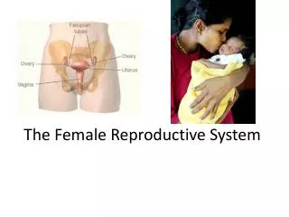
The Female Reproductive System. Figure 26–20c The Appearance of the Endometrium during the Uterine Cycle. The Female Reproductive System. Blood Supply of the Vagina Is through vaginal branches of internal iliac (uterine) arteries and veins. The Female Reproductive System.

E N D
The Female Reproductive System Figure 26–20c The Appearance of the Endometrium during the Uterine Cycle.
The Female Reproductive System • Blood Supply of the Vagina • Is through vaginal branches of internal iliac (uterine) arteries and veins
The Female Reproductive System • Innervation of the Vagina • Hypogastric plexus • Sacral nerves • Branches of pudendal nerve
The Female Reproductive System • Vaginal Lamina Propria • Is thick and elastic • Contains small blood vessels, nerves, and lymph nodes
The Female Reproductive System • The Vaginal Mucosa • Is surrounded by elastic muscularis layer • Layers of smooth muscle fibers • Arranged in circular and longitudinal bundles • Continuous with uterine myometrium
The Female Reproductive System • The Vaginal Epithelium • Is nonkeratinized, stratified, and squamous • Forms folds (rugae) • Changes with ovarian cycle
The Female Reproductive System • Vulva (or pudendum) • Area containing female external genitalia • Vestibule • A central space bounded by small folds (labia minora) • Covered with smooth, hairless skin • Urethra opens into vestibule • Anterior to vaginal entrance
The Female Reproductive System • Paraurethral Glands • Also called Skene glands • Discharge into urethra near external opening
The Female Reproductive System • Mammary Glands • Consist of lobes • Each containing several secretory lobules • Separated by dense connective tissue
The Female Reproductive System • Suspensory Ligaments of the Breast • Bands of connective tissue • Originate in dermis of overlying skin • Areolar tissue separates mammary gland complex from underlying pectoralis muscles • Blood Supply of Mammary Glands • Branches of internal thoracic artery
The Female Reproductive System • Innervation of the Vagina • Hypogastric plexus • Sacral nerves • Branches of pudendal nerve
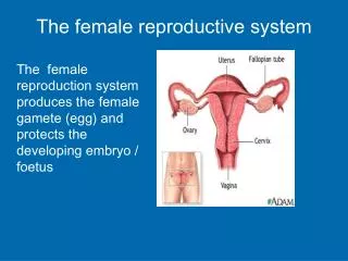
The Female Reproductive System • Three Functions of the Vagina • Passageway for elimination of menstrual fluids • Receives spermatozoa during sexual intercourse • Forms inferior portion of birth canal
The Female Reproductive System • The Vaginal Wall • Contains a network of blood vessels and layers of smooth muscle • Is moistened by • Secretions of cervical glands • Water movement across permeable epithelium
The Female Reproductive System • The Hymen • Is an elastic epithelial fold • That partially blocks entrance to vagina • Ruptured by sexual intercourse or tampon usage
The Female Reproductive System • Vaginal Muscles • Two bulbospongiosus muscles extend along either side of vaginal entrance • Vestibular bulbs: • masses of erectile tissue that lie beneath the muscles • have same embryological origins as corpus spongiosum of penis
The Female Reproductive System • The Vaginal Epithelium • Is nonkeratinized, stratified, and squamous • Forms folds (rugae) • Changes with ovarian cycle
The Female Reproductive System • Vaginal Lamina Propria • Is thick and elastic • Contains small blood vessels, nerves, and lymph nodes
The Female Reproductive System • The Vaginal Mucosa • Is surrounded by elastic muscularis layer • Layers of smooth muscle fibers • Arranged in circular and longitudinal bundles • Continuous with uterine myometrium
The Female Reproductive System Figure 26–21 The Histology of the Vagina.
The Female Reproductive System • Vaginal Bacteria • A population of harmless resident bacteria • Supported by nutrients in cervical mucus • Creates acidic environment • Restricts growth of many pathogens
The Female Reproductive System • A Vaginal Smear • Is a sample of epithelial cells shed at surface of vagina • Used to estimate stage in ovarian and uterine cycles
The Female Reproductive System • Vulva (or pudendum) • Area containing female external genitalia • Vestibule • A central space bounded by small folds (labia minora) • Covered with smooth, hairless skin • Urethra opens into vestibule • Anterior to vaginal entrance
The Female Reproductive System • Menses • Is the degeneration of functional zone • Occurs in patches • Is caused by constriction of spiral arteries • Reducing blood flow, oxygen, and nutrients • Weakened arterial walls rupture • Releasing blood into connective tissues of functional zone
The Female Reproductive System • Three Suspensory Ligaments of Uterus • Uterosacral ligaments • Prevent inferior–anterior movement • Round ligaments • Restrict posterior movement • Cardinal(lateral) ligaments • Prevent inferior movement
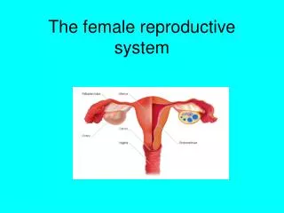
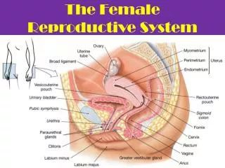
The Female Reproductive System • Uterine Body (or corpus) • Is largest portion of uterus • Ends at isthmus • Fundus • Is rounded portion of uterine body • Superior to attachment of uterine tubes
The Female Reproductive System • Cervix • Is inferior portion of uterus • Extends from isthmus to vagina • Distal end projects about 1.25 cm (0.5 in.) into vagina • External os • Also called external orifice of uterus • Is surrounded by distal end of cervix • Leads into cervical canal
The Female Reproductive System • Cervical Canal • Is a constricted passageway opening to uterine cavity of body • At internal os (internal orifice)
The Female Reproductive System • Blood Supply of the Uterus • Branches of uterine arteries • Arising from branches of internal iliac arteries • Ovarian arteries • Arising from abdominal aorta • Veins and lymphatic vessels
The Female Reproductive System • Nerves of the Uterus • Autonomic fibers from hypogastric plexus (sympathetic) • Sacral segments S3 and S4 (parasympathetic) • Segmental blocks • Anesthetic procedure used during labor • Target spinal nerves T10–L1
The Female Reproductive System Figure 26–18a The Uterus.
The Female Reproductive System Figure 26–18b The Uterus.
The Female Reproductive System • The Uterine Wall • Has a thick, outer, muscular myometrium • Has a thin, inner, glandular endometrium (mucosa)
The Female Reproductive System • Two Divisions of Endometrium • Functional zone • Layer closest to uterine cavity • Basilar zone • Adjacent to myometrium
The Female Reproductive System • Menses • Is the degeneration of functional zone • Occurs in patches • Is caused by constriction of spiral arteries • Reducing blood flow, oxygen, and nutrients • Weakened arterial walls rupture • Releasing blood into connective tissues of functional zone
The Female Reproductive System Figure 26–20b The Appearance of the Endometrium during the Uterine Cycle.
The Female Reproductive System • The Secretory Phase • Endometrial glands enlarge, increasing rate of secretion • Arteries of uterine wall • Elongate and spiral through functional zone • Begins at ovulation and persists as long as corpus luteum remains intact • Peaks about 12 days after ovulation • Glandular activity declines • Generally lasts 14 days
The Female Reproductive System • The Vagina • Cervix projects into vaginal canal • Fornix is shallow recess surrounding cervical protrusion • Lies parallel to • Rectum, posteriorly • Urethra, anteriorly
The Female Reproductive System • Blood Supply of the Vagina • Is through vaginal branches of internal iliac (uterine) arteries and veins
The Female Reproductive System • The Secretory Phase • Endometrial glands enlarge, increasing rate of secretion • Arteries of uterine wall • Elongate and spiral through functional zone • Begins at ovulation and persists as long as corpus luteum remains intact • Peaks about 12 days after ovulation • Glandular activity declines • Generally lasts 14 days
The Female Reproductive System Figure 26–20c The Appearance of the Endometrium during the Uterine Cycle.
The Female Reproductive System • The Uterine Cycle • Ends as corpus luteum stops producing stimulatory hormones
The Female Reproductive System • Menarche • The first uterine cycle • Begins at puberty (age 11–12) • Menopause • The termination of uterine cycles • Age 45–55
The Female Reproductive System • Amenorrhea • Primary amenorrhea • Failure to initiate menses • Transient secondary amenorrhea • Interruption of 6 months or more • Caused by physical or emotional stresses
The Female Reproductive System • The Vagina • Is an elastic, muscular tube • Extends between cervix and vestibule • 7.5–9 cm (3-3.6 in.) long • Highly distensible
The Female Reproductive System • The Vagina • Cervix projects into vaginal canal • Fornix is shallow recess surrounding cervical protrusion • Lies parallel to • Rectum, posteriorly • Urethra, anteriorly
The Female Reproductive System • Blood Supply of the Vagina • Is through vaginal branches of internal iliac (uterine) arteries and veins
The Female Reproductive System • Menses • Is the degeneration of functional zone • Occurs in patches • Is caused by constriction of spiral arteries • Reducing blood flow, oxygen, and nutrients • Weakened arterial walls rupture • Releasing blood into connective tissues of functional zone
The Female Reproductive System • Three Suspensory Ligaments of Uterus • Uterosacral ligaments • Prevent inferior–anterior movement • Round ligaments • Restrict posterior movement • Cardinal(lateral) ligaments • Prevent inferior movement
The Female Reproductive System • Two Divisions of Endometrium • Functional zone • Layer closest to uterine cavity • Basilar zone • Adjacent to myometrium