Download
1 / 32
320 likes | 434 Views
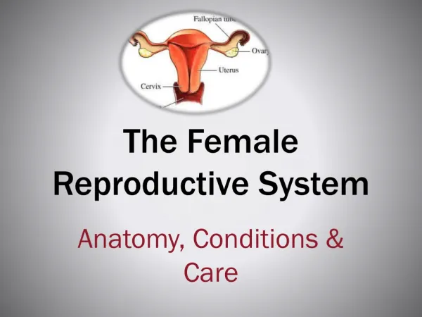
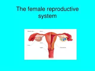
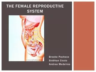
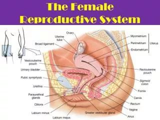
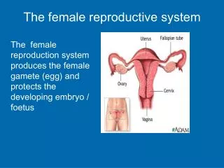
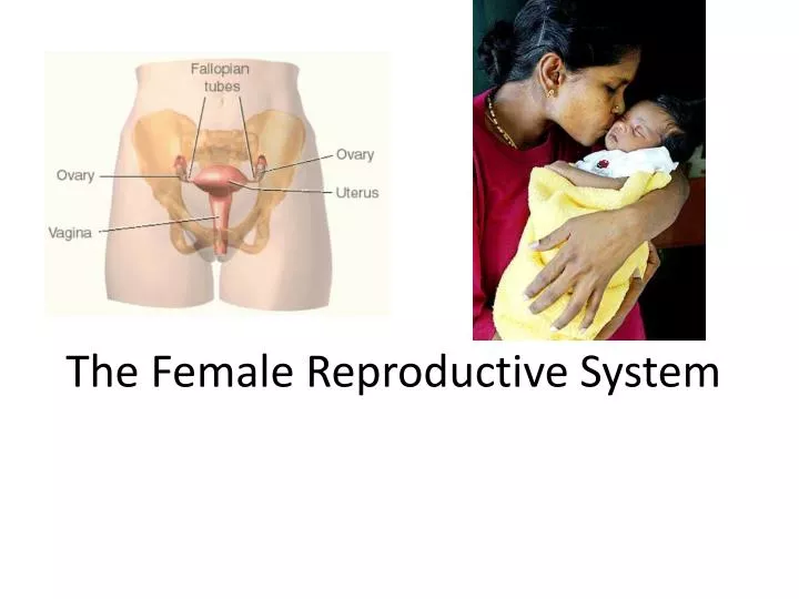
The Female Reproductive System. Female Reproductive Structures. Female Reproductive Structures. Step by Step – The life of a female. Ovaries develop in ___________ Produce sex hormones (___________ and ___________) 400 000 ___________ develop (potential eggs)

E N D
Step by Step – The life of a female • Ovaries develop in ___________ • Produce sex hormones (___________ and ___________) • 400 000 ___________ develop (potential eggs) • Constant ___________ after puberty • Each menstrual cycle, ~________ follicles develop but only one becomes mature
Do the math! Reproductive success declines after age 27 Question: How many periods will a female have in her life? • ______________ Question: How long will she be able to reproduce? (Assuming her first period is at age 13) • ___________ ___________ ___________
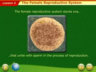
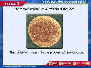
Skip to Puberty: Oogenesis Ovary • ___________ • Follicles have two kinds of cells • Primary ___________( ___________ becomes the egg) • ___________ cells (nutrient for the ___________) • ______from pituitary causes follicle development • _____= ______________________________________
___________ • Nutrient cells around primary ___________ divide • ___________ I (in the ovary) • Majority of ___________ to one cell (___________ oocyte) • ________body produced • ___________ II (occurs later if it contacts a sperm) • Majority of ___________ to one cell again (egg) • ___________ body produced
Ovulation Cells around primary ___________ create a fluid filled cavity ___________ pushes outwards ___________ vessels weaken Secondary ___________ released = ___________
Ovulation • Surrounding ___________ cells become the ___________ ___________ • Produces ___________ to prepare uterus • No pregnancy? • ___________ ___________ dies after _____ days
Secondary ___________ enters the ___________ (___________ tube) Carried by ___________ Could be ___________ here If it doesn’t meet a ___________ , the oocyte will die within ___________ hours Takes ___________ days to travel to the uterus (___________ from the ___________ ___________ stops uterine contractions!)
Fertilization (maybe?) Secondary ___________ and sperm could meet in ___________ (oocyte egg) ___________ on sperm breaks into the egg Only one sperm can enter
Fertilized Egg development (maybe?) • Fertilized egg (zygote) begins to divide
Implantation (maybe?) ___________ _____ can implant in the ___________ (nourishes the egg) Implantation in the ______ = _______ (___________ pregnancy)
If fertilization DOESN’T occur (likely!) • ___________ ___________ eventually dies • Lack of ___________ causes uterine contracts • ___________! • Sheds ___________ • Start all over again…
Animal Development Umbilical blood vessels Mammalian embryo Chorion Bird embryo Amnion Yolk sac Allantois Fetal blood vessels Placenta Maternal blood vessels
Human fetal development 10 weeks
Placenta • ___________ exchange across membranes
Human embryonic development • embryo showing ___________ & ___________ buds
Human embryonic development • beginning of the eye can be seen, as well the bulging ___________ & the ___________ ___________
Human embryonic development • ___________ of the eye can be seen forming, the mass of the ___________ bulging from the chest, & the beginnings of the ___________ rays
Human embryonic development • beginning of the ___________ is clearly seen note the bend of the elbow joint has begun, the fingers are forming and toes are beginning to bud off the foot
Human embryonic development • Note the formation of the ___________ , ___________ , ear flap & well defined toes & fingers 50–60 days (8 weeks) Both knee & elbow are visible. Embryo has formed most of basic organ systems & will spend remainder of development in “fetal” period. Organs grow, mature, & begin to learn their respective functions
Human fetal development 4 weeks 7 weeks
Human fetal development • Week 9 - week 40 = ___________ • after ____ weeks or so, the baby's development is largely "finished" • some exceptions: ___________ & ___________ development Week 16
Human fetal development 12 weeks
Human fetal development • The ___________ just spends much of the 2nd & 3rd trimesters just growing Week 20
Human fetal development • 24 weeks (6 months; 2nd trimester) fetus is covered with fine, downy hair called lanugo. Its skin is protected by a waxy material called vernix
Human fetal development • 30 weeks (7.5 months) umbilical cord
Getting crowded in there!! • 32 weeks (8 months) The fetus sleeps 90-95% of the day & sometimes experiences REM sleep, an indication of dreaming
Birth (36 weeks) Intestine Placenta Umbilical cord Wall of uterus Bladder Cervix Vagina