Download
1 / 24
680 likes | 1.59k Views
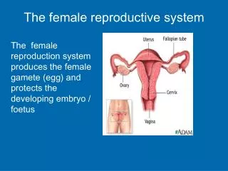
Histology of the Female Reproductive System. PROF. DR. FAUZIAH OTHMAN DEPT OF HUMAN ANATOMY. Histology of ovaries. Ovary consist of the following part; Germinal epithelium =simple epithelium( low cuboidal or squamous) cover ovary.

E N D
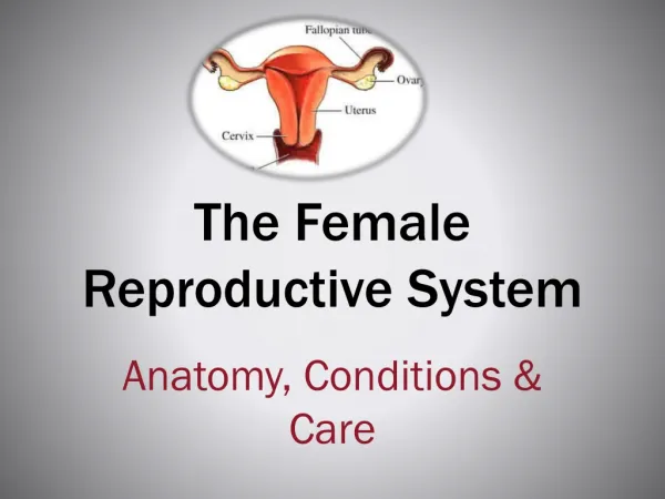
Histology of the Female Reproductive System PROF. DR. FAUZIAH OTHMAN DEPT OF HUMAN ANATOMY
Ovary consist of the following part; • Germinal epithelium =simple epithelium( low cuboidal or squamous) cover ovary. • Tunica albuginea = whitish capsule of dense irregular connective tissue located immediately deep to the germinal epithelium • Ovarian medulla = deep to ovarian cortex. The medulla consists of more loosely arranged connective tissue & contains blood vessels, lymphatic vessels & nerves.
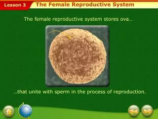
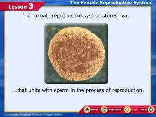
Ovarian follicle = in cortex & consist of oocytes in various stage of development, plus the cell surrounding them. • When surrounding cells • form a single layer –follicular cells. • Several layer- Granulosa cells
Primordial follicles – located in the cortex below tunica albuginea. • Single layer of squamous follicular cells. • Primary follicles – change to cuboidal or low columnar cells. • Developing oocytes –have a large eccentric nucleus with a conspicuous nucleolus. • By mitosis from cuboidal cells called granulosa cells. • Single layer of granulosa cells surround the oocyte form the corona radiata.
Bet corona radiata and oocyte, a noncellular glycoprotein layer called zona pellucida. • Stromal cells surround the follicular cells differentiate into theca interna. • Thin bm separates granulosa cells from the theca interna cells. • Atretic follicle – degenerating primordial, developing, or mature follicles. • Secondary follicle – during growth of follicles, fluid accumulates bet granulosa cells – antrum.
Corpus luteum -collapsed folded mass of glandular epithelium consists primarily of theca lutein and granulosalutein cells. • Large cells, have large vesicular nuclei, and stain lightly because of lipid inclusions. • Theca lutein cells (former theca interna cells) are external to the granulosa lutein cells on the periphery of the glandular epithelium. • Smaller than granulosa cells , cytoplasm stains darker, and smaller and darker. • If ovulated oocyte is not fertilized, corpus luteum regress to scar tissue known as corpus albicans. • Anticipation of the implantation of a fertilized egg and pregnancy. • Lutein cells secrete level of estrogen and progesterone to stimulate development of uterus and mammary glands. • Theca lutein cells extends inward with the CT septa into folds. • Theca externa cells forms a poorly defined capsule around the corpus luteum and inward with the CT septa into the fold
Wall of uterus • Perimetrium= serosa/ adventitia • Myometrium= thick smooth muscle • Endometrium = simple epithelium that descend into a lamina propria to form numerous uterine glands.
Endometrium • 2 functional layer • A) luminal stratum functionalis • B) stratum basalis • Non-pregnant- during menstrual, superficial of functionalis is slought off, leaving intact the deeper basalis layer with the basal remnant of the uterine glands
Lining epithelium Lamina propria Functionalis layer Coiled arteries Basalis layer Uterine glands Smooth muscle (longitudinal) Interstitial connective tissue Smooth muscle (cross section) Blood vesels Histology of uterus
Columnar epithelium Uterine gland with secretion Functionalis layer Lamina propria (with edema) Coiled arteries Uterine glands (hypertrophied & tortous) Basalis layer Blood vessel Smooth muscle bundles Myometrium
Uterine tube Simple columnar overlying the vascularized & loose connective tissue lamina propria. Luminal epithelium consist 2 cell-ciliated cells & nonciliated peg cell. 2 smooth muscle- circular muscle (inner) & longitudinal muscle (outer) Serosa- outermost layer on uterine tubes
Vagina • Lumen- stratified squamous epithelium • Lamina propria- contains small blood vessels, lymphatic tissue, dense irregular connective tissue • Muscularis of vaginal wall- longitudinal bundles & oblique bundles of smooth muscle • Adventitia – blood vessel & nerves bundles.
Cervix • Cervical canal-tall, mucus-secreting columnar epithelium • Lamina propria- more fibrous than uterus