Download
1 / 25
330 likes | 779 Views
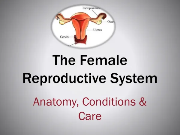
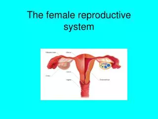
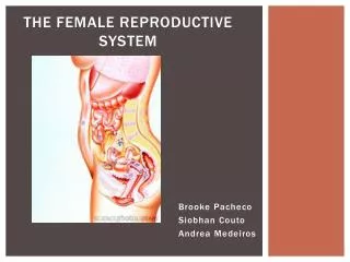
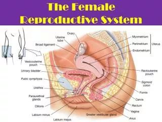
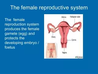
The Female Reproductive System. By Dr D Fisher. Anatomy of Female Reproductive System. Female reproductive organs Ovaries Uterine tubes Uterus Vagina External genital organs Mammary glands. Female Reproductive Anatomy. Female Pelvis.

E N D
The Female Reproductive System By Dr D Fisher
Anatomy of Female Reproductive System • Female reproductive organs • Ovaries • Uterine tubes • Uterus • Vagina • External genital organs • Mammary glands
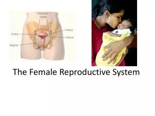
Uterus, Vagina, Uterine Tubes, Ovaries and Supporting Ligaments
The Developing Follicle • Atresia of all follicles but one • One follicle develops faster than the rest • Starts to secrete estrogens: • Enhances granulosa cell proliferation • Increase no. of FSH receptors on granulosa cells • Increase FSH effect on follicle • Decrease secretion of FSH & LH
Oogenesis is the production of a secondary oocyte in ovaries Oogonia are cells from which oocytes develop Primary oocytes are surround by granulosa cells and called a primordial follicle Primordial follicle becomes a primary follicle when oocyteenlarges and cells change Primary follicle becomes secondary follicle and enlarges to form mature or graafian follicle Usually only one is ovulated, others degenerate Primary oocyte completes first meiotic division to produce secondary oocyte and a polar body Secondary oocyte begins second meiotic division, which stops in metaphase II Follicle and Oocyte Development
Ovulation Follicle swells and ruptures, secondary oocyte is released from ovary Second meiotic division completed when secondary oocyte unites with sperm cell to form zygote Fate of the follicle Graafian follicle become corpus luteum If fertilization occurs, corpus luteum persists If no fertilization, becomes corpus albicans Ovulation and Follicle Fate
Functions of FSH • Stimulates growth and maturation of ovarian follicles: Primary to mature Graafian follicle. • Theca interna: secretion of estrogen (follicular stage) • Granulosa cells: Secretion of progesterone (luteal stage)
Functions of LH • Initially supports the stimulatory effect of FSH. • Imperative for ovulation to occur (day 14). • LH causes the granulosa cells to form lutein cells (luteinization) which forms the corpus luteum. • Lutein cells secrete estrogen and progesterone. • Lutein cells: proliferation→enlargement →secretion →degeneration.
Puberty Begins with menarche or first episode of menstrual bleeding Begins when GnRH levels increase Menstrual Cycle About 28 days long Phases Menses Proliferative phase Secretory phase Menses Amenorrhea: Absence of a menstrual cycle Menopause: Cessation of menstrual cycles Puberty and Menstrual Cycle
Proliferative Phase (estrogen phase) • Increase concentration of estrogens secreted by ovaries. • Stromal and epithelial cells proliferate rapidly. • Increase growth of glands & B/Vs into endometrium. • At the time of ovulation→endometrium 3mm thick.
Secretory Phase (progesterone Phase) • Increase secretions of estrogen and progesterone from the corpus luteum. • Progesterone causes: • Marked swelling of the endometrium. • Increased secretory development. • Increase nutritional content of stromal cells • Increase gland tortutosity • Endometrium thickness: 6mm.
Menstrual Phase • Day 26 → degeneration of corpus luteum → • decrease levels of blood estrogen and progesterone: • Decrease stimulation of endometrial cells • Involution of endometrium (by 65%). • Bld vessels become vasoplastic (constrict) • Necrosis of endometrium • All superficial layers of endometrium desquamated (broken down) • Uterine wall contracts →expels contents: • 35ml blood • 35ml serous fluid • Ps.: Fibrolysin prevents clotting