Download
1 / 27
280 likes | 792 Views
INTRODUCTION. The spinal cord and spinal nerves mediate reactions to environmental changes.The spinal cord has several functions.It processes reflexes.It is the site for integration of EPSPs and IPSPs that arise locally or are triggered by nerve impulses from the periphery and brain.It is a cond

E N D
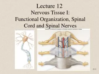
1. Chapter 13: The Spinal Cord & Spinal Nerves
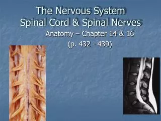
2. INTRODUCTION The spinal cord and spinal nerves mediate reactions to environmental changes.
The spinal cord has several functions.
It processes reflexes.
It is the site for integration of EPSPs and IPSPs that arise locally or are triggered by nerve impulses from the periphery and brain.
It is a conduction pathway for sensory and motor nerve impulses.
The size of the vertebral canal varies in different regions of the vertebral column and determines seriousness of spinal cord injuries.
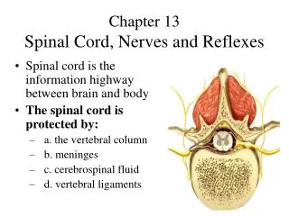
3. Spinal cord anatomy: protective structures Vertebral column
Spinal cord is located within the vertebral cavity
Layer of fat and connective tissue in epidural space between vertebra and meninges.
Meninges�3 connective tissue covers that are continuous with the cranial meninges
4. Spinal cord anatomy: protective structures
5. Meninges Dura mater forms a sac from the foramen magnum to S2.
Subdural space has interstitial fluid.
Arachnoid mater has spider�s web arrangement of delicate collagen fibers.
Subarachnoid space has cerebral spinal fluid.
Pia mater is a thin transparent connective tissue that covers the spinal cord.
Has blood vessels, collagen and elastic fibers
Denticulate ligaments are thickenings of pia mater that fuse with arachnoid mater and dura mater between anterior and posterior nerve roots
6. Meninges
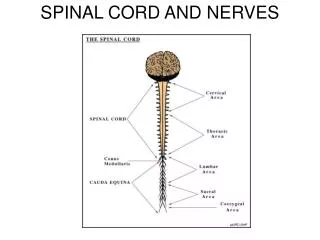
7. External anatomy Spinal cord extends from the medulla oblongata to super border of L2.
In newborns it extends to L3 or L4.
Growth stops at 5 years
Cervical and lumbar enlargement
Conus medullaris
Cauda equina
Filum terminale�extension of pia mater that anchors spinal cord to coccyx
8. Spinal nerves Each pair arises from a spinal segment
Spinal nerves are named for segment where they are located.
Bundles of axons called roots connect the spinal nerves to the spinal cord.
Dorsal root has sensory axons
Dorsal root ganglion has sensory cell bodies
Ventral root has motor axons
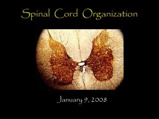
9. Spinal cord: internal anatomy
10. Spinal nerves Are part of the PNS.
Connect the CNS to sensory receptors, muscles, and glands in the body.
NOT ALL SPINAL CORD ROOTS ARE ALIGNED WITH CORRESPONDING VERTEBRAE.
Have two roots�anterior is motor and posterior is sensory.
The two roots join to make up the nerve that is classified as a mixed nerve.
11. Spinal nerve coverings Individual axons are wrapped in endoneurium
Groups of axons are arranged in fascicles and are wrapped in perineurium.
The entire nerve is wrapped in epineurium.
Dura mater fuses with epineurium of the spinal nerves as they pass through the intervertebral foramen.
12. Spinal nerve coverings
13. Distribution of spinal nerves After passing through the intervertebral foramen the spinal nerve branches.
Branches are called rami (singular, ramus).
Anterior ramus serves the muscles and structures of the upper and lower limbs, and skin of lateral and ventral surfaces.
14. Spinal nerve branches Posterior ramus serves deep muscles and skin of dorsal trunk.
Meningeal branch reenters the intervertebral foramen and supplies all structures in the vertebral column.
Rami communicantes are components of the ANS.
15. Plexuses Axons from anterior rami form networks on both sides of the body with axons from adjacent nerves.
A network of axons is called a plexus.
Cervical, brachial, lumbar, sacral, and lumbar plexus
Anterior rami of T2-T12 are called intercostal or thoracic nerves and do not form plexuses.
16. Dermatomes Definition: the area of skin that provides sensory information to one pair of spinal nerves.
Damaged regions of spinal cord can be identified by stimulating dermatome regions
17. Spinal cord physiology The white matter contains sensory and motor tracts.
The gray matter contains cell bodies and integration sites for EPSPs and IPSPs
Spinal nerves connect the CNS to sensory receptors, muscles, and glands.
18. Reflexes Reflex is a fast, automatic, unplanned sequence of actions that occur in response to a particular action.
Inborn reflexes
Acquired reflexes
Reflexes produced by integration in the spinal cord are spinal reflexes.
Reflexes produced by integration in the brain stem are cranial reflexes.
Somatic reflexes involve muscular contractions
Autonomic reflexes involve responses of cardiac and smooth muscle and glands.
19. Reflex arc
20. Reflex arc Sensory receptor
Sensory neuron
Integrating center
Monosynaptic reflex arc has one synapse in the CNS
Polysynaptic reflex arc involves more than 2 types of neurons and CNS synapses.
Motor neuron
Effector
Somatic reflex
Autonomic or visceral reflex
21. Somatic spinal reflexes Stretch reflex�causes contraction of a skeletal muscle in response to stretching of the muscle.
Tendon reflex�a feedback mechanism to control muscle tension by causing muscle relaxation (protects against torn tendons)
Flexor or withdrawal reflex�an ipsilateral reflex (intersegmental reflex arc)
Crossed extensor reflex�a contralateral reflex that synchronizes extension of contralateral limb.
22. The stretch reflex
23. Muscle spindle and tendon organ
24. Stretch reflex Stretch reflex pathway is ipsilateral and can be either mono or polysynaptic.
The simultaneous contraction of agonistic muscle and relaxation of antagonistic muscle is termed reciprocal innervation.
Reciprocal innervation prevents conflict between opposing muscle groups and promotes coordination of muscle groups.
Stretch reflex also helps maintain posture
25. Tendon reflex
26. Flexor or withdrawal reflex
27. Flexor and crossed extensor reflex
28. Reflexes and diagnoses Patellar reflex
Achilles reflex or ankle jerk
Babinski sign and negative Babinski sign
Abdominal reflex
Pupillary light reflex Patellar reflex�extension of legs at the knee joint by contraction of quadriceps femoris in response to tapping the patellar ligament. Reflex is absent in those with diabetes mellitus or neurosyphilis and exaggerated in those who have disease or injury to particular motor tracts
Achilles reflex or ankle jerk�a stretch reflex that involves extension (plantar flexion) of the foot by contraction of gastocnemius and soleus muscle in response to tapping the calcaneous. Reflex is also affected by diabetes, neurosyphylis, alcoholism and subarachnoid hemorrhages. Compression of cervical cord or lesion of tracts in sacral area can exaggerate response.
Babinski sign and negative Babinski sign- produced by stroking lateral margin of the sole. The large toe dorsiflexes and there is lateral fanning of the other toes. Indication of incomplete myelination most commonly seen in children under 1.5 yrs. A negative Babinski sign is a curling under of the toes
Abdominal reflex�contraction of the muscles that compress abdominal wall in response to stroking the side of the abdomen. The muscle contraction causes the umbilicus to move in the direction of the stimulus. Absence indicates problems with peripheral nerves, lesions of integrating centers or MS.
Pupillary light reflex�response to light. Absence may indicate brain damage or injury.
Tortora and Derrickson, 2005Patellar reflex�extension of legs at the knee joint by contraction of quadriceps femoris in response to tapping the patellar ligament. Reflex is absent in those with diabetes mellitus or neurosyphilis and exaggerated in those who have disease or injury to particular motor tracts
Achilles reflex or ankle jerk�a stretch reflex that involves extension (plantar flexion) of the foot by contraction of gastocnemius and soleus muscle in response to tapping the calcaneous. Reflex is also affected by diabetes, neurosyphylis, alcoholism and subarachnoid hemorrhages. Compression of cervical cord or lesion of tracts in sacral area can exaggerate response.
Babinski sign and negative Babinski sign- produced by stroking lateral margin of the sole. The large toe dorsiflexes and there is lateral fanning of the other toes. Indication of incomplete myelination most commonly seen in children under 1.5 yrs. A negative Babinski sign is a curling under of the toes
Abdominal reflex�contraction of the muscles that compress abdominal wall in response to stroking the side of the abdomen. The muscle contraction causes the umbilicus to move in the direction of the stimulus. Absence indicates problems with peripheral nerves, lesions of integrating centers or MS.
Pupillary light reflex�response to light. Absence may indicate brain damage or injury.
Tortora and Derrickson, 2005