Download
1 / 15
160 likes | 398 Views
Nervous Tissue I: Functional Organization, Spinal Cord and Spinal Nerves. Lecture 12. Nervous Tissue. Found in brain, spinal cord and nerves Property Ability to produce action potentials (electric signals) Cells Nerve cells or neurons Neuroglia or support cells

E N D
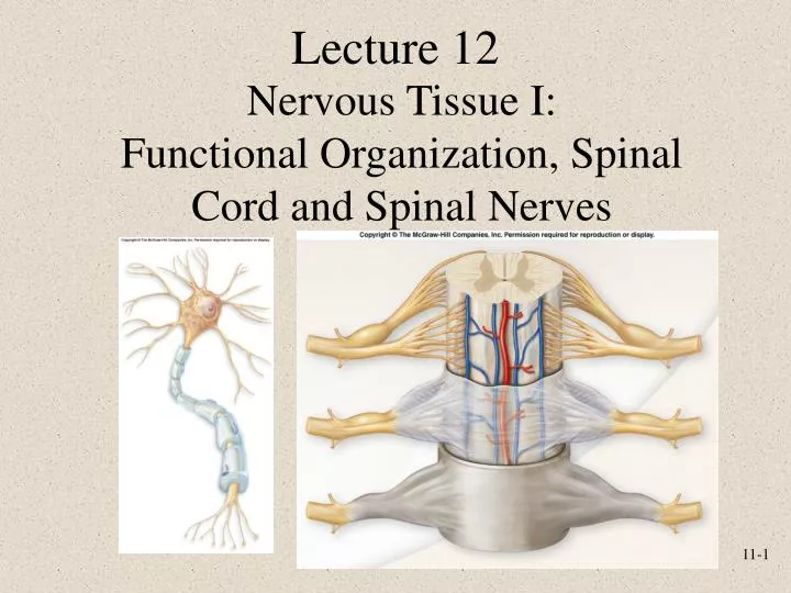
Nervous Tissue I:Functional Organization, Spinal Cord and Spinal Nerves Lecture 12
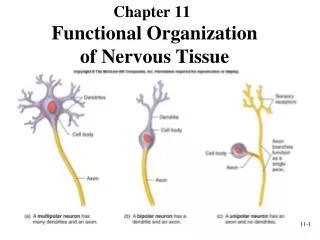
Nervous Tissue • Found in brain, spinal cord and nerves • Property • Ability to produce action potentials (electric signals) • Cells • Nerve cells or neurons • Neuroglia or support cells • Oligodendrocytes and Schwann cells
The Nervous System • Subdivisions • Central nervous system (CNS) • Peripheral nervous system (PNS) • Sensory receptor • Receptor of sensory information • Nerve • Made up of a bundle of axons • Ganglion • Collection of cell bodies of neurons • Plexus • Network of spinal nerves
Central Nervous System • Consists of • Brain • Located in cranial vault of skull • Spinal cord • Located in vertebral canal • Brain and spinal cord • Continuous with each other at foramen magnum • Tract Fig. 14.1
Peripheral Nervous System • Two subcategories • Sensory or afferent • Motor or efferent • Divisions • Somatic nervous system • Autonomic nervous system (ANS) • Sympathetic (fight or flight) • Parasympathetic (rest and digest) Fig. 14.2
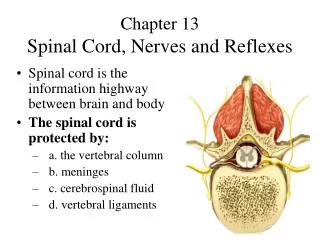
Spinal Cord • Extends from foramen magnum to second lumbar vertebra • Segmented • Cervical • Thoracic • Lumbar • Sacral • Gives rise to 31 pairs of spinal nerves • Not uniform in diameter throughout length Fig. 16.1
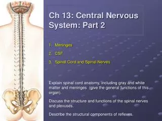
Meninges • Connective tissue membranes surrounding spinal cord and brain • Dura mater • Arachnoid mater • Pia mater • Spaces • Epidural: Anesthesia injected • Subdural: Serous fluid • Subarachnoid: cerebrospinal fluid (CSF) Fig. 16.2
Cross Section of Spinal Cord • White matter • Myelinated axons forming tracts • Three funiculi (columns) • Gray matter • Neuron cell bodies, dendrites, axons • Three horns Fig. 16.3 Fig. 16.4
Spinal NervesCervical Plexus • C1-C4 • Phrenic nerve • from C3-C5 (cervical and brachial plexus) • innervates diaphragm Fig. 16.8
Brachial Plexus • C5-T1 • Major nerves • Radial • Ulnar • Median Fig. 16.9
Lumbar Plexus Fig. 16.10
Sacral Plexus Fig. 16.11
Review Question Compression of the ________ nerve against the medial epicondyle of the humerus will produce strong tingling sensations along the forearm and hand. Radial Median Phrenic Femoral Ulnar
Points to Remember • Nervous system consists of central nervous system (brain and spinal cord) and peripheral nervous system (all nervous tissue outside of central nervous system) • Sensory (afferent) neurons carry sensory information to brain and spinal cord • Motor (efferent) neurons carry motor away from brain and spinal cord to spinal nerves and cranial nerves • Spinal nerves have a dorsal root (sensory neurons) and a ventral root (motor neurons) • Names of nerves in plexuses generally describe the body region they travel