Download
1 / 68
740 likes | 2.36k Views
What are the major components of a spinal nerve?. Spinal Nerves. Figure 13–6. Organization of Spinal Nerves. Every spinal cord segment: is connected to a pair of spinal nerves Every spinal nerve: is surrounded by 3 connective tissue layers that support structures and contain blood vessels.

E N D
Spinal Nerves Figure 13–6
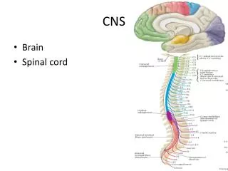
Organization of Spinal Nerves • Every spinal cord segment: • is connected to a pair of spinal nerves • Every spinal nerve: • is surrounded by 3 connective tissue layers • that support structures and contain blood vessels
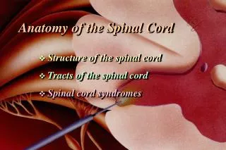
3 Connective Tissue Layers • Epineurium: • outermost layer • dense network of collagen fibers • Perineurium: • middle layer • divides nerve into fascicles (axon bundles) • Endoneurium: • inner layer • surrounds individual axons
Peripheral Nerves • Interconnecting branches of spinal nerves • Surrounded by connective tissue sheaths
How does the distribution pattern of spinal nerves relate to the regions they innervate?
Peripheral Distribution of Spinal Nerves • Spinal nerves: • form lateral to intervertebral foramen • where dorsal and ventral roots unite • then branch and form pathways to destination
Nerve Plexuses • Contain no synapses! • For pre-midterm (Summarized in tables in text and lab guide): • Know cord roots (“ventral rami,” actually) that contribute to the plexus • Know the names of the major peripheral nerves that each plexus gives rise to. 3D Rotation of Peripheral Nerves and Nerve Plexuses PLAY Figure 13–9
The Cervical Plexus Figure 13–10
The Lumbar and Sacral Plexuses • Innervate pelvic girdle and lower limbs 3D Rotation of Lumbar and Sacral Plexuses PLAY Figure 13–12a, b
The Lumbar and Sacral Plexuses Figure 13–12c, d
Post-Viral inflammation of the sensory nerves Rash follows dermatomes. Notice it does not cross the midline. Medical Example: Shingles
Dermatomes • Bilateral region of skin • Monitored by specific pair of spinal nerves Figure 13–8
Peripheral Distribution of Spinal Nerves • Sensory fibers Figure 13–7b
Peripheral Distribution of Spinal Nerves • Motor fibers PLAY Peripheral Distribution of Spinal Nerves Figure 13–7a
Functional Organization of Neurons • Sensory neurons: • about 10 million • deliver information to CNS • Motor neurons: • about 1/2 million • deliver commands to peripheral effectors
Functional Organization of Neurons • Interneurons: • about 20 billion • interpret, plan, and coordinate signals in and out • often organized into functional “neuronal pools”
5 Patterns of Neural Circuits in Neuronal Pools • Divergence: • spreads stimulation to many neurons or neuronal pools in CNS Figure 13–13a
5 Patterns of Neural Circuits in Neuronal Pools • Convergence: • brings input from many sources to single neuron Figure 13–13b
5 Patterns of Neural Circuits in Neuronal Pools • Serial processing: • moves information in single line Figure 13–13c
5 Patterns of Neural Circuits in Neuronal Pools • Parallel processing: • moves same information along several paths simultaneously Figure 13–13d
5 Patterns of Neural Circuits in Neuronal Pools • Reverberation: • positive feedback mechanism • functions until inhibited Figure 13–13e
Development of Reflexes • A reflex is a rapid, predictable motor response to a stimulus. • Innate reflexes are unlearned and involuntary • Acquired reflexes are complex, learned motor patterns
Nature of Reflex Responses • Somatic: Reflexes involving skeletal muscles and somatic motor neurons. • Autonomic (visceral) Reflexes controlled by autonomic neurons • Heart rate, respiration, digestion, urination, etc • Spinal reflexes are integrated within the spinal cord gray matter while cranial reflexes are integrated in the brain. • Reflexes may be monosynaptic or polysynaptic
Components of a Reflex Arc • 1. Activation of a Receptor: site of stimulus • 2. Activation of a Sensory Neuron: transmits the afferent impulse to spinal cord (CNS) • 3. Information processing at the Integration center: synapses (monosynaptic reflexes) or interneurons (polysynaptic) between the sensory and motor neurons. • In CNS • Spinal reflexes or cranial reflexes
Components of a Reflex Arc • 4. Activation of a Motor Neuron: transmits the efferent impulse to effector organ • 5. Response of a peripheral Effector: Muscle or gland that responds
Spinal Reflexes • 4 important somatic spinal reflexes • Stretch • Tendon • Flexor(withdrawal) • Crossed extensor reflexes
Stretch Reflexes • 1. Stretching of the muscle activates a muscle spindle (receptor) • 2. An impulse is transmitted by afferent fibers to the spinal cord • 3. Motor neurons in the spinal cord cause the stretched muscle to contract • 4. The integration area in the spinal cord • Polysynaptic reflex arc to antagonist muscle causing it to to relax (reciprocal innervation)
Notice hammer Stretch Reflex
Stretch Reflex ExamplePatellar Reflex • Tap the patellar tendon • muscle spindle signals stretch of muscle • motor neuron activated & muscle contracts • Quadriceps muscle contracts • Hamstring muscle is inhibited (relaxes) • Reciprocal innervation (polysynaptic- interneuron) • antagonistic muscles relax as part of reflex • Lower leg kicks forward • Demonstrates sensory and motor connections between muscle and spinal cord are intact.
Tendon Reflexes • Monitors external tension produced during muscular contraction to prevent tendon damage • Controls muscle tension by causing muscle relaxation • Golgi tendon organs in tendon (sensory receptor) • activated by stretching of tendon • inhibitory neuron is stimulated • motor neuron is hyperpolarized and muscle relaxes • Both tendon & muscle are protected • Reciprocal innervation (polysynaptic) • causes contraction Martini pg 443 states the receptor is unidentified; this is incorrect.
Notice no hammer Tendon Reflex
Flexor Reflex • Withdrawal reflex • When pain receptors are activated it causes automatic withdrawal of the threatened body part.
Flexor (Withdrawal) Reflex • Is this a monosynaptic or a polysynaptic reflex? • Is this an ipsilateral or a contralateral reflex?
Crossed Extensor Reflex • Complex reflex that consists of an ipsilateral withdrawal reflex and a contralateral extensor reflex • This keeps you from falling over, for example if you step on something painful. When you pull your foot back, the other leg responds to hold you up.
Superficial Reflexes • Elicited by gentle cutaneous stimulation • Important because they involve upper motor pathways (brain) in addition to spinal cord neurons
Superficial ReflexesPlantar Reflex • Tests spinal cord from L4 to S2 • Indirectly determines if the corticospinal tracts of the brain are working • Draw a blunt object downward along the lateral aspect of the plantar surface (sole of foot) • Normal: Downward flexion (curling) of toes
Plantar Reflex Normal Abnormal (Babinski’s)
Abnormal Plantar Reflex: Babinski’s Sign • Great toe dorsiflexes (points up) and the smaller toes fan laterally • Happens if the primary motor cortex or corticospinal tract is damaged • Normal in infants up to one year old because their nervous system is not completely myelinated.
Organization Similarities of SNS and ANS Figure 16–2
Visceral Reflexes • Provide automatic motor responses • Can be modified, facilitated, or inhibited by higher centers, especially hypothalamus
Visceral Reflexes Figure 16–11
Case of the Woman with HT • Name the two parts of the ANS • Describe the two major groups of receptors and their subtypes (and their usual ligands.) • Distinguish between receptor stimulation and cell stimulation. • Explain what “specificity” means when we are referring to a ligand’s specificity for receptors. • Provide a background for studying examples of somatic and autonomic reflexes.
Nerve Plexuses • Complex, interwoven networks of nerve fibers • Formed from blended fibers of ventral rami of adjacent spinal nerves • Control skeletal muscles of the neck and limbs