Download
1 / 29
430 likes | 1.52k Views
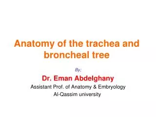
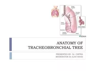
ANATOMY OF TRACHEOBRONCHIAL TREE. PRESENTED BY- Dr. CHITRA MODERATOR- Dr.AJAY SOOD. Respiratory System. TRACHEA. It is a cartilaginous and membranous tube EXTENT - from 6 th cervical vertebra till the body of 5 th thoracic vertebra

E N D
ANATOMY OF TRACHEOBRONCHIAL TREE PRESENTED BY- Dr. CHITRA MODERATOR-Dr.AJAY SOOD
TRACHEA It is a cartilaginous and membranous tube • EXTENT - from 6th cervical vertebra till the body of 5th thoracic vertebra • During expiration the bifurcation rises by one vertebral level and during inspiration may be lowered as far as 6th thoracic vertebra • LENGTH - 11cm • DIAMETER - 2 to 2.5cm • CHILDREN - smaller, deeper, more moveable
STRUCTURE • CARTILAGES-16to20 in no.,each forms an incomplete ring,which occupies anterior two third of circumference of trachea • Are placed horizontally above each other,separated by narrow intervals • 4mm deep and 1mm thick • Outer surface is flattened in vertical direction and convex from inner side • Highly elastic,but may calcify in later stages • FIRST TRACHEAL CARTILAGE-broader,divided connected to lower end of cricoid by cricotracheal ligament • LAST TRACHEAL CARTILAGE-thick and broad in midlle, lower border is prolonged to a triangular hook shaped process which curves downward and backward between two bronchi
FIBROUS MEMBRANE-cartilages are enclosed in an elastic fibrous membrane which consists of two layers,one passes over the outer surface the other one over the inner surface • At upper and lower margins they blend together to form a single membrane • MUSCULAR TISSUES-two layers of non –striated muscles longitudinal and transeverse • Longitudinal fibres are external,consist of few scattered bundles only • Transeverse fibers(trachealis muscle) are internal,extends between the end of cartilages
MUCUS MEMBRANE-continous above with larynx and below with bronchus • Consist of areolar and lymphoid tissue,basementmembrane,supporting stratified epithelium ,surface layer of which is columnar and ciliated • Beneath basement membrane there is a layer of longitudinal elastic fibre • Submucuslayer,composed of loose meshwork of connective tissue VESSELS AND NERVES- • ARTERIAL SUPPLY-Inferior thyroid arteries • NERVE SUPPLY-vagusnerve,recurrentnerve,sympathetic nerves
RIGHT BRONCHUS • 2.5cm long • Wider,shorter,more vertical • Divides into EPARTERIAL and HYPARTERIAL BRONCHUS based on right pulmonary artery
RIGHT UPPER LOBE BRONCHUS • It divides into three segmental bronchi which supply apical,anterior and posterior segments of upper lobe • APICAL SEGMENTAL BRONCHUS-divides into apical and anterior subsegmental branches • POSTERIOR SEGMENTAL BRONCHUS-divides into lateral and anterior subsegmental branches. It serves the posteroinferior part of superior lobe of lung • ANTERIOR SEGMENTAL BRONCHUS-runs anteroinferiorly to supply rest of the part of upper bronchus. Divides into lateral and anterior subsegmental branches
RIGHT MIDDLE LOBE BRONCHUS • Divides into lateral and medial subsegments RIGHT LOWER LOBE BRONCHUS • Continuation of principal stem beyond the origin of middle lobe bronchus • Supplies 5 segments of the lung • Apical segmental bronchus • Medial basal segmental bronchus • Anterior basal segmental bronchus • Lateral and posterior basal segmental bronchus
LEFT BRONCHUS • 5cm in length • Smaller in caliber • Enters the lung opposite 6th thoracic vertebra • RELATIONS-passes beneath the aortic arch,crosses in front of oesophagus,thoracicduct,descendingaorta,has left pulmonary artery at first above and then in front
LEFT UPPER LOBE BRONCHUS • Cranially it divides into anterior ,apical and posterior segmental branches • Caudally into superior and inferior lingual branches LEFT LOWER LOBE BRONCHUS • Divides into apical segmental bronchus,medialbasal,anteriorbasal,lateral and posterior basal branches
BRONCHI • - • Right bronchus • Wider • More vertical • shorter • Supported by C shaped cartilages • 20-30 degree angle • First generation • Left bronchus • Narrower • More angular • Longer • Supported by C shaped cartilages • 40-60 degree angle • First generation
CLINICAL SIGNIFICANCE • Right main bronchus is more in line with trachea • Inhaled foreign bodies and gastric contents enter right bronchial tree • If patient is lying on his side,lateralsubsegments of anterior and posterior segments are more likely to get such material • If patient is supine,then apical segmental bronchus which arise from right or left lower lobe bronchus is the most common part of lung for the aspirated material to collect
BRONCHOPULMONARY SEGMENTS • Anatomic,functional and surgical units of lung • 23 divisons or generations are involved in dichotomous division,starting from trachea till alveolar sacs • Each lobar bronchus(secondary bronchus) gives off branches called segmental bronchus(tertiary bronchus)
As bronchi become smaller, cartilages also become smaller and fewer in no • Bronchioles are formed which are less then 1mm in dia,no cartilages and lined by ciliated columnar epithelium • Divide to form terminal bronchioles,which show delicate outpouchings from their wall • These are respiratory bronchioles,dia is 0.5mm,they end by branching into alveolar ducts
Each alveolar sac consist of around 17 alveoli • Each alveolus surrounded by rich network of blood capillaries • Gas exchange primarily occurs on thin side of alveolocapillarymembrane,thick side provides structural support
ALVEOLI • Type I pneumocytes-large flattened cells, present a very thin diffusion barrier for gases • Type II pneumocytes-secretes surfactant,which decreases the surface tension between thin alveolar walls • macrophages
Subdivision of lung lobe • Pyramid shaped,apex towards lung root • Surrounded by connective tissue • Segmental bronchus,segmentalartery,lymph vessels and autonomic nerves • Segmental vein lies between adjacent segments
BLOOD SUPPLY OF LUNGS • By bronchial arteries which are branches of descending aorta NERVE SUPPLY OF LUNGS • Pulmonary plexus-efferent and afferent autonomic nerve fibres • Sympathetic efferent fibres produce bronchodilation and vasoconstriction • Parasympathetic efferent produces bronchoconstriction,vasodilation,increase glandular secretions