Download
1 / 10
100 likes | 256 Views
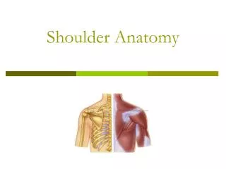
Anatomy of Shoulder. Bones: Clavicle. Slender S-shaped Supports the anterior portion of the shoulder Keeping it free from the thoracic cage Extends from the sternum to tip of shoulder where it j oins the acromion process of scapula. Bones: Clavicle. Medial 2/3 is circular

E N D
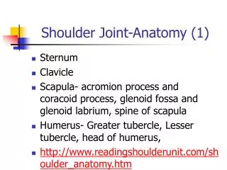
Bones: Clavicle • Slender • S-shaped • Supports the anterior portion of the shoulder • Keeping it free from the thoracic cage • Extends from the sternum to tip of shoulder where it joins the acromion process of scapula.
Bones: Clavicle • Medial 2/3 is circular • Lateral third more flattened • Medial 2/3 bend convexly forward • Lateral 3rd is concave • Pt. at which the clavicle changes shape and contour causes weakness • Large number of fx occur at that point
Bones: Sternum • Also known as breastbone • 3 parts • manubrium • Body • Xiphoid process • Attachment • Clavicle at SC joint • Which is the only axial skeleton attachment for the entire upper extremity
Bones: Scapula • Flat, triangular shaped bone • Serves mainly as an articulating surface for the head of the humerus • 3 prominent projections • Spine • Acromion • Coracoid process • Site of attachment for many muscles that act to move the shoulder complex
Bones: Humerus • Head: spherical with a shallow constricted neck • Articulating with the scapula’s glenoid fossa • Greater and lesser tubercles • Lesser: positioned anteriorly and medially • Greater: lateral • Between the two tubercles is the bicipital groove • Long tendon of the biceps brachii muscle
Joints: Sternoclavicular Joint • Clavicle articulates with the manubrium of sternum • Fibrocartilaginous disk is a shock absorber against medial forces and prevents any displacement upward
Joints: Acromioclavicular Joint • Gliding articulation of the lateral end of the clavicle with acromion process • Weak junction • Thin fibrous capsule surrounds the joint
Joints: Glenohumeral Joint • Ball and socket joint • Cavity is deepened slightly by the fibrocartlaginous rim called the glenoid labrum • Active and passive mechanisms • Passive: glenoid labrum and capsular ligaments • Active: deltoid and rotator cuff muscles
Joints: Scapulothoracic Joint • Not a true joint • Movement of the scaupla on the wall of the thoracic cage is critical to shoulder joint movement • Allows for scapular elevation, depression, protraction, retraction, abduction and adduction