Download
1 / 30
310 likes | 555 Views
Eukaryotic Viruses. Very small- can only be detected by electron microscopy or by indirect clinical analysis. Obligate intracellular parasites: viruses cannot replicate without the help of a host cell Contents nucleic acid (DNA or RNA) a few proteins—to help it establish infection

E N D
Eukaryotic Viruses Very small- can only be detected by electron microscopy or by indirect clinical analysis. Obligate intracellular parasites: viruses cannot replicate without the help of a host cell Contents nucleic acid (DNA or RNA) a few proteins—to help it establish infection capsid—proteinaceous coat lipid coating taken from host cell (in some cases)
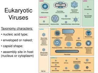
Classification of viruses The nucleic acids they carry (DNA vs. RNA) The nature of the capsid surrounding the virus (icosahedral vs. helical) Whether they are naked—containing only DNA/RNA and capsid encapsulated –also surrounded by a lipid bilayer derived from a host cell.
Classifications of viruses based on nucleic acid, capsid and presence or absence of envelope ** **
RNA viruses Serves as mRNA can be directly translated into viral proteins! Must be converted to + RNA first RETROVIRUSES CYTOPLASM NUCLEUS nucleus cytoplasm
DNA Viruses nucleus cytoplasm
Capsid—protein coat thatsurrounds DNA or RNA Shapes of viruses ICOSAHEDRAL RNA or DNA viruses HELICAL always an RNA virus
Icosahedral symmetry capsid Form globular protein from polypetide chain (3o structure) Arrange globular proteins Into equilateral triangle Place twenty triangles together to form icosahedron
Helical capsid symmetry for RNA viruses Capsomer—small protein subunits associated with RNA like beads on a string When RNA forms a helical structure, the capsomer proteins are able to form one large helical capsid as they interact with each other
Viruses can be naked or encapsulated This depends on how the virus leaves the host cell it has previously infected Naked—If virus reaches critical mass and causes the cell to burst (similar to P1 bacterial phage we discussed) Encapsulated—If virus buds out of the cell taking some of the lipid bilayer from that cell.
When virus buds from nucleus or cell membrane it can take host lipid bilayer with it—such virus is encasulated as opposed to naked
Typical life cycle of virus 1. Make contact with host cell—usually specific 2. Bind to a receptor on the cell surface 3. Enter the cell via endocytosis or fusion of membranes 4. Uncoat the virus to reveal the nucleic acids RNA virus –cytoplasm DNA and retroviruses must enter nucleus first 5. Translate mRNA or + stranded RNA that acts like mRNA 6. Make proteins required for structural proteins proteins responsible for RNA synthesis 7. Exit cell to infect other cells and spread misery.
Life cycle of RNA viruses adsorption and uptake n.b. virus inside of cell now coated with lipid, this must be removed n.b. virus inside of cell no longer covered with lipid Naked virus Encapsulated protein
Replication of positive stranded RNA viruses • + RNA immediately translated • into protein required to make • RNA and + RNA (rdRNAP) • The - RNA is replicated via • rdRNAP to make lots of +RNA • The +RNA is translated to make • Coat proteins (capsid) original +RNA GAUCGAUCG -RNA CUAGCUAGC +RNA GAUCGAUCG template progeny
Replication of negative stranded RNA viruses • RNA enters cell with • its own vitral RNAP that • converts – RNA into • + RNA • +RNA translated to make • progeny – RNA and • capsid
Influenza VirusClass Orthomyxovirussegmented – stranded RNA virushelical capsidenveloped Indications: Fever/chills muscle and joint aches, headache stomach ache and cold-like symptoms Influenza Virus A infects humans, swine and birds, most likely to cause Flu Pandemics Influenza Virus B and C only isolated from humans, causes Flu epidemics
Influenza virus—upper respiratory tract cells M protein (matrix protein) tethers HA and NA to lipid bilayer of virus NA (neuraminidase) binds to mucin and cleaves the neuramic acid that makes up mucin. Reveals the sialic acid receptor HA (Hemaglutinase) binds to sialic acid receptors of host cell. Once binding established, virus can fuse with host cell.
Why do we suffer from the Flu if we have had it before?Antigenic Drift Our body makes antibodies to HA and NA BUT during replication of viral RNA small changes are made in the HA and NA genes. point mutations small deletions This changes the antigenic nature of the HA and NA proteins such that our body doesn’t recognize these proteins and MUST mount a new immune response N.B. Flu is usually self limiting even though the HA and Na has been changed the change may be small enough that we can mount a weak immune response. Mild symptoms
Why do we see Flu pandemics that can kill a large number of people? Antigenic Shift 1918—Spanish Flu, killed up to 40 million people worldwide 1957—Asian Flu, low mortality 1968—Hong Kong Flu, low mortality, avian flu virus the poultry was destroyed. Antigenic Shift leads to a complete change in the NA and/or HA!!! --2 different influenza viruses attack the same animal --The RNA of the flu virus is segmented such that different RNA segments from different sources can be packaged IN 2 known viruses OUT 2 new viruses
Treatment and control Treatment DO NOT give aspirin to children, aspirin causes Reye’s Syndrome (severe liver and brain pathology) Amanatadine or Rimantidine –prevents uncoating of Influenza Virus A Sanamavir (inhaled) oseltamivar (oral) –neuraminidase inhibitors, neuraminidase cannot break down mucin Prevention Vaccines. Scientists choose 3 strains circulating in a population and grow these in chick embryos. Virus isolated, inactivated purified and used to make vaccines Vaccines given to elderly, immuno-compromised and health care workers.
Retroviruses can transform normal cells into tumor cells by introducing or activating oncogenes Oncogenes gene that causes uncontrolled growth of cells 1.carried into cells certain retroviruses (leukemia, sarcomas) 2. Can be present in humans as proto-oncogenes oncogenes that are inactive unless a. A carcinogen mutates a region near the gene b. A retrovirus inserts near the proto-oncogene
Acute transforming virus with oncogenes • Binds to a receptor • Taken in by fusion • of lipids • Retroviral RNA converted • to DNA in the cytoplasm via • reverse transcriptase • DNA enters nucleus • DNA integrates into • chromosome viaintegrase • (much like a transposon) • DNA transcribed into • large mRNA molecule • Large protein translated • Viral proteases cleave • large inactive protein into • smaller active proteins Viral oncogene inserted into chromosme
The product of oncogenes leads to uncontrolled cell growth Cell surface receptors that bind to mitogens. Mitogens induce Phosphorylation of tyrosine residues on receptors signaling for normal cell growth and division Similar to EGF, PDGF and insulin receptor only has MORE tyrosine kinaseactivity!!!
A retrovirus can integrate near an inactive protooncogene and activate that gene’s expression Retrovirus contains promoters that up-regulate the expression of the proto-oncogene that normally does not have its own promoter!!
HIV and AIDS Usually causes acute disease HIV can integrate into the host chromosome and lie dormant for years Activation? Stimulation of T-cells by an infection from something else may lead to transcription of the quiet integrated virus. HIV kills T helper cells HIV can evade the immune system by travelling from one T-cell to another without leaving the cell
HIV Structure Gp160 (glycoprotein) binds to CD4 receptors found on T helper cells Enters cells with a lot of its own proteins: reverse transcriptase, integrase and proteases
HIV genes Sticky ends recognized by integrase reverse transcriptase and envelop genes mutate at a high rate.
HIV Vaccines • Vaccines to the gp160 protein • 1. prevents gp160 from binding to CD4 receptors on T-cells • Caveats • 1. gp160 changes at a high rate • 2. HIV can “hide” from antibodies by traveling • from cell to cell without leaving cell • Give patients high dosages of CD4 receptors: HIV binds to • the exogenous receptors instead of to T helper cells
HIV treatment Prevent reverse transcriptase activity or protease activity. • NRTI’s (nucleoside reverse transcriptase inhibitors) • AZT resembles deoxy-thymidine, therefore as the virus converts • its RNA to DNA it inserts AZT in lieu of dT. DNA elongation • aborted. • NNRTI’s (non- nucleoside reverse transcriptase inhibitors) • binds to rev. transcriptase thus inactivating it. • Protease inhibitors—computer designed peptide analogs that binds to • protease. Large inactive protein not cleaved into smaller active • proteins. • Usually a combination of all classes of drugs given as virus mutates at a high rate