Download
1 / 37
370 likes | 508 Views
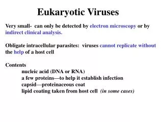
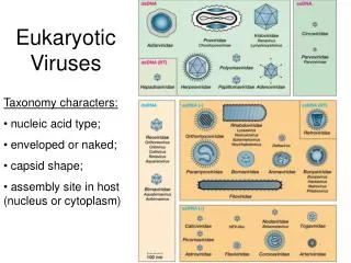
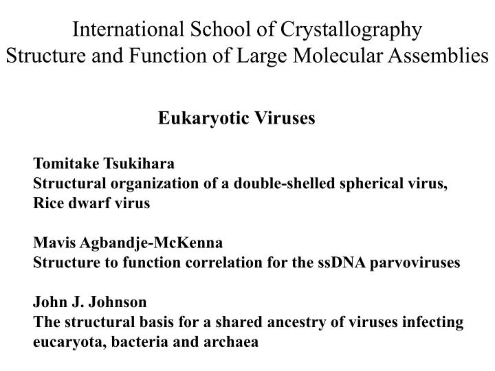
International School of Crystallography Structure and Function of Large Molecular Assemblies. Eukaryotic Viruses. Tomitake Tsukihara Structural organization of a double-shelled spherical virus, Rice dwarf virus Mavis Agbandje-McKenna

E N D
International School of Crystallography Structure and Function of Large Molecular Assemblies Eukaryotic Viruses Tomitake Tsukihara Structural organization of a double-shelled spherical virus, Rice dwarf virus Mavis Agbandje-McKenna Structure to function correlation for the ssDNA parvoviruses John J. Johnson The structural basis for a shared ancestry of viruses infecting eucaryota, bacteria and archaea
Structure organization of a double-shelled spherical virus, Rice dwarf virus T. TsukiharaIPR, Osaka University, Japan
Hierarchy of assembly Accurate inter-molecular recognition Motivation Why are such complicated structures as RDV, cytochrome c oxidase, eukaryotic proteasome assembled correctly in the cell?
Double stranded RNA Viruses Family: Reoviridae 12 genome segments 10 genome segments 10 genome segments Reovirus (Reinisch et al., 2000) RDV (Nakagawa et al., 2003) BTV (Grimes et al., 1998) RDV (Nakagawa et al., 2003)
Rice Dwarf Virus (RDV) Guanylyltransferase Non structural proteins Pns4 Pns6 Pns10 Pns11 Pns12
Diffraction data and structure refinement of RDV Space group: I222 Cell constants (Å): a=770, b=795, c=814 Resolution (Å) : 230.0-3.5 Total film packs: 1477 Observed reflections: 17,806,888 Unique reflections: 3,001,937 Completeness(%): 97.7 Rmerge: 0.186 Rcryst: 0.303 Rfree: 0.306 r.m.s. deviation bond lengths 0.010 Å bond angles 1.48° Ramachandran plots most favored region 85.2% disallowed region 0.1%
Electron density map of RDV refined by NCS averaging Contour level is 1.0σ 50 Å resolution 25 Å resolution
BTV P1,polymerase P5,guanylyl transferase P7,RNA binding protein Transcription complex (Polymerase & Capping enzyme) dsRNA BTV (Gouet et al., Cell, 1999) P8 P3 RNA
The electron density of P7 pentamer. RDV P7(289~300 SEPFSDKERSEL) P7(55KDa), an RNA binding protein directly interact with P3.
Inner core Outer shell
Structure comparison of the inner capsid proteins of Reoviridae Reovirus l1A RDV P3A RDV P3B BTV VP3A
P3 subunit structure 90 P3A 90 P3B
E G S F R K Intermolecular interactions G-F, strongest interaction --> Dimer 2. Circular E-F interaction --> Decamer *Number of other atom pairs with distances less than 4Å
The P3-decamer accepts a transcription complex and a genome segment in the viral inclusion. Five P7 molecules combine with P3 decamers around five-fold axis. (Nakagawa et al., Structure, 2003) P7 was included in a P3 core, when both proteins were co-expressed. ( Hagiwara et al., 2003, JGV) A viral RNA tightly interacts with P1, P5 and P7 during the structural organization. (Zhong et al., Science China, 2004) P1, P3, P5 and P7 are in the viral inclusion consisting of Pns6, Pns11 and Pns12. Viral RNAs are synthesized in the viral inclusion. (Wei et al., 2006, JGV)
300 nm Pns11 Pns12 Pns6 Immunogold labelling of electron-dense inclusions with Pns6, Pns11 and Pns12. Dark dots indicate these three proteins. The viral inclusions consist of these non-structural proteins. (Wei et al., 2006, JGV)
P1 The inner core proteins, P1, P3, P5 and P7 coexist with Pns12 in viral inclusions. P3 P5 P7 P2 The outer shell proteins, P2, P8 and P9 are not in the viral inclusions. P8 P9 Confocal fluorescence microscopy (Wei et al., 2006, JGV)
The core particles were obtained by incubating virus particles in 1.4M MgCl solution. (Takahashi & Omura et al., 1994) P3 can form the inner core without P8.
Simplified animation of assembly process of the inner core in the viral inclusion
Proposed model for structure organization ofRDV particle (1) (Decamer model)
Structure organization of the outer shell P8 trimers Nucleation of trimers on the three-fold axes of the inner core. Two dimensional growth on the inner core surface
P8 subunit structure The P8 trimer is rigid.
P P P E E E Q Q Q R R R G G G F F F S S S S S S T T T R R R L L L K K K P8 trimer - inner core interaction *Number of other atom pairs with distances less than 4Å
0.8 M MgCl2 treated Wu & Omura et al., 2000
Zhu & Omura et al., 1997 P8-trimers tend to make a two-dimensional hexagonal array.
Assembly of P8-trimers on the inner core surface
The symmetry mismatch between the inner core and the outer shell is overcome by the sequential assembly following the nucleation.
Summary Hierarchy of structural organization is achieved by protein sorting in the viral inclusion and ranking of inter-molecular interactions. It reduces the freedom of assembly process. Symmetry enable viruses to assemble by a few kinds of inter-molecular interactions. Accurate inter-molecular recognition is achieved by accurate inter-atomic interactions.